Copyright
©2006 Baishideng Publishing Group Co.
World J Gastroenterol. Jun 21, 2006; 12(23): 3779-3781
Published online Jun 21, 2006. doi: 10.3748/wjg.v12.i23.3779
Published online Jun 21, 2006. doi: 10.3748/wjg.v12.i23.3779
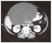
Figure 1 Contrast-enhanced CT scan demonstrates a large liver cyst in the left liver lobe (arrow).
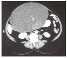
Figure 2 CT scan shows the cyst with contrast-enhanced locules (arrow).
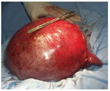
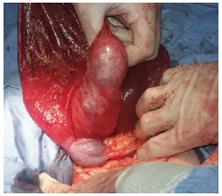
Figure 3 Intra-operative view of the large liver cyst.
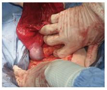
Figure 4 Intra-operative aspects after puncture of the tumor.
The gallbladder lies over the cyst’s wall.
Figure 5 An area with increased consistency and irregular surface in the posterior region of the tumor.
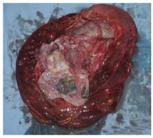
Figure 6 The surgical specimen with multiple locules and septations.
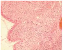
Figure 7 Photo-micrograph de-monstrates a columnar epithe-lium and an “ovarian-like” stroma (HE staining, × 200).
- Citation: Beuran M, Venter MD, Dumitru L. Large mucinous biliary cystadenoma with “ovarian-like” stroma: A case report. World J Gastroenterol 2006; 12(23): 3779-3781
- URL: https://www.wjgnet.com/1007-9327/full/v12/i23/3779.htm
- DOI: https://dx.doi.org/10.3748/wjg.v12.i23.3779


