Copyright
©2006 Baishideng Publishing Group Co.
World J Gastroenterol. Jun 21, 2006; 12(23): 3776-3778
Published online Jun 21, 2006. doi: 10.3748/wjg.v12.i23.3776
Published online Jun 21, 2006. doi: 10.3748/wjg.v12.i23.3776
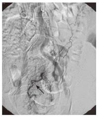
Figure 1 Arteriogram picture taken before the 1st embolization.
Arrow demonstrates an abnormal vasculature.
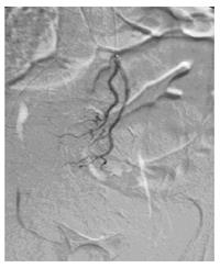
Figure 2 Arteriogram picture taken immediately after the 1st embolization.
No abnormal vessels were seen.
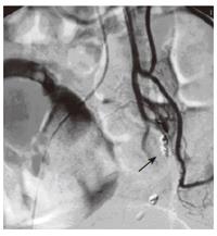
Figure 3 Arteriogram picture taken after the 2nd embolization.
Arrow demonstrates the Gel foam pledget and metallic coil at the superior rectal artery.
- Citation: Yip VS, Downey M, Teo NB, Anderson JR. Management of ischemic proctitis with severe rectal haemorrhage: A case report. World J Gastroenterol 2006; 12(23): 3776-3778
- URL: https://www.wjgnet.com/1007-9327/full/v12/i23/3776.htm
- DOI: https://dx.doi.org/10.3748/wjg.v12.i23.3776


