Copyright
©2006 Baishideng Publishing Group Co.
World J Gastroenterol. Jun 14, 2006; 12(22): 3581-3584
Published online Jun 14, 2006. doi: 10.3748/wjg.v12.i22.3581
Published online Jun 14, 2006. doi: 10.3748/wjg.v12.i22.3581
Figure 1 Morphology change of apoptosis in HT-29 cells induced by C2-ceramide at different concentrations for different time under transmission electron microscope.
A: 0 μmol/L, 24 h, × 5000; B: 50 μmol/L, 24 h, × 4000; C: 50 μmol/L, 48 h, × 3000.
Figure 2 Morphology change of apoptosis in HT-29 cells induced by C2-ceramide at different concentrations for different time under fluorescence microscope.
A: 0 μmol/L, 24 h, × 100; B: 50 μmol/L, 12 h, × 100; C: 50 μmol/L, 24 h, × 100).
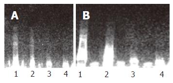
Figure 3 DNA fragmentation induced by C2-ceramide in HT-29 cells for 12 h (A) and 24 h (B).
1: 50 μmol/L; 2: 25 μmol/L; 3: 12.5 μmol/L; 4: 0 μmol/L.
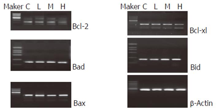
Figure 4 Expression of apoptosis-related genes in HT-29 cells induced by C2-ceramide for 24 h (C: 0 μmol/L; L: 12.
5 μmol/L; M: 25 μmol/L; H: 50 μmol/L).
- Citation: Zhang XF, Li BX, Dong CY, Ren R. Apoptosis of human colon carcinoma HT-29 cells induced by ceramide. World J Gastroenterol 2006; 12(22): 3581-3584
- URL: https://www.wjgnet.com/1007-9327/full/v12/i22/3581.htm
- DOI: https://dx.doi.org/10.3748/wjg.v12.i22.3581


