Copyright
©2006 Baishideng Publishing Group Co.
World J Gastroenterol. Jun 14, 2006; 12(22): 3466-3470
Published online Jun 14, 2006. doi: 10.3748/wjg.v12.i22.3466
Published online Jun 14, 2006. doi: 10.3748/wjg.v12.i22.3466
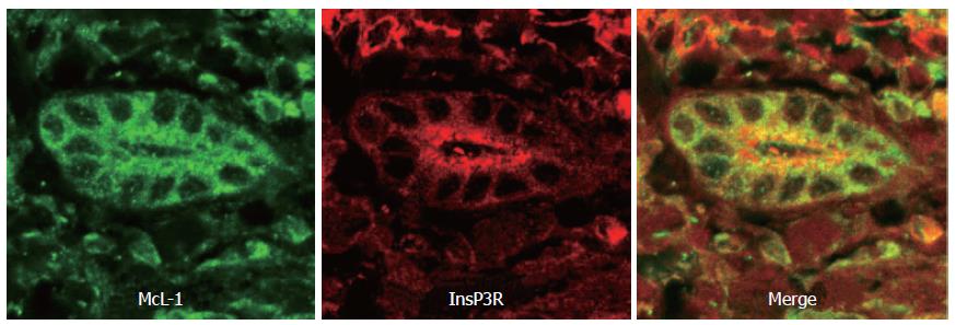
Figure 1 Distribution of the InsP3 receptor and Mcl-1 in human bile ducts.
This confocal immunofluorescence image was obtained from a paraffin-embedded biopsy specimen from a normal liver. The specimen was double-labeled with antibodies against Mcl-1 (green) and the type III InsP3R (red). Note that the Mcl-1 is distributed diffusely throughout the cytosol, while the InsP3R is most concentrated in the apical region.
- Citation: Minagawa N, Ehrlich BE, Nathanson MH. Calcium signaling in cholangiocytes. World J Gastroenterol 2006; 12(22): 3466-3470
- URL: https://www.wjgnet.com/1007-9327/full/v12/i22/3466.htm
- DOI: https://dx.doi.org/10.3748/wjg.v12.i22.3466


