Copyright
©2006 Baishideng Publishing Group Co.
World J Gastroenterol. May 21, 2006; 12(19): 3077-3081
Published online May 21, 2006. doi: 10.3748/wjg.v12.i19.3077
Published online May 21, 2006. doi: 10.3748/wjg.v12.i19.3077
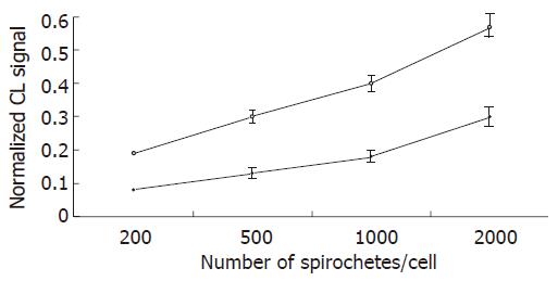
Figure 1 Chemiluminescence (CL) detection of reactive oxygen species generation by Kupffer cells stimulated by L.
interrogans and B.burgdorferi. Kupffer cells were incubated in triplicate with borrelia (white circles) and leptospira (black circles) preparations at various bacteria/KC ratio, for 3 h in the presence of 0.6 mM luminol. The chemiluminescence emissions were acquired at 2 min intervals for 3 h. The experiments were repeated three times, and data points represent the mean ± SD of the integrated chemiluminescence signal, normalized to that of the control.
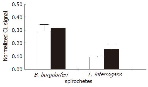
Figure 2 Effect of spirochete opsonization on the chemiluminescence (CL) signal.
Each value represents the mean ± SD of three independent experiments carried out at a ratio of 500 spirochetes/KCs using unopsonized (white bars) and opsonized (grey bars) bacteria. Note that the ROS production induced by borreliae was about twice the ROS generation of leptospirae.
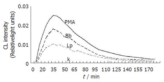
Figure 3 Chemiluminescence kinetic profiles of reactive oxygen radical generation in Kupffer cells treated with L.
interrogans and B.burgdorferi. Bacteria were used at a ratio of 2000/KCs. Lp and Bb stand for leptospirae and borreliae, respectively. PMA represents Kupffer cells treated with phorbol myristate acetate, whereas K represents untreated control Kupffer cells.
Figure 4 iNOS determination by SDS-polyacrylamide gel electrophoresis and by Western blotting in Kupffer cells stimulated by L.
interogans and B. burgdorferi. The 130 ku iNOS protein band was detected by specific polyclonal rabbit anti-iNOS antibodies. The iNOS protein band was evident 2 h after infection. The band peaked 6 h after infection and was still evident 22 h after infection. Lane 1: Kupffer cells stimulated by LPS and interferon-γ (positive control); lane 2: Kupffer cells not stimulated (negative control); lane 3: Kupffer cells stimulated by L.interrogans; lane 4: Kupffer cells stimulated by B.burgdorferi.
- Citation: Marangoni A, Accardo S, Aldini R, Guardigli M, Cavrini F, Sambri V, Montagnani M, Roda A, Cevenini R. Production of reactive oxygen species and expression of inducible nitric oxide synthase in rat isolated Kupffer cells stimulated by Leptospira interrogans and Borrelia burgdorferi. World J Gastroenterol 2006; 12(19): 3077-3081
- URL: https://www.wjgnet.com/1007-9327/full/v12/i19/3077.htm
- DOI: https://dx.doi.org/10.3748/wjg.v12.i19.3077


