Copyright
©2006 Baishideng Publishing Group Co.
World J Gastroenterol. May 21, 2006; 12(19): 3065-3072
Published online May 21, 2006. doi: 10.3748/wjg.v12.i19.3065
Published online May 21, 2006. doi: 10.3748/wjg.v12.i19.3065
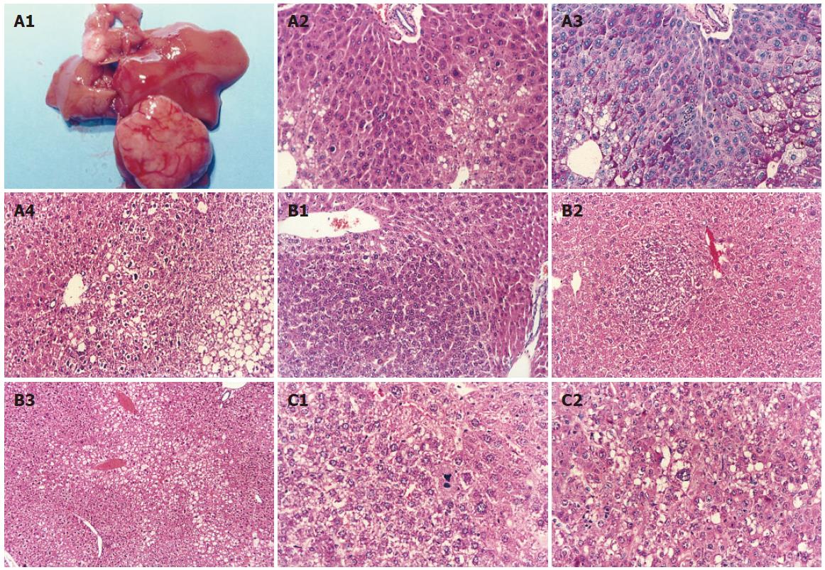
Figure 1 Histopathological alterations identified in liver tissues of mice treated with either AFB1 (A) and cyanobacterial toxins alone (B) or in combination (C).
A1: Grossly identified hepatocelluar carcinoma (HCC) in an HBVx gene transgenic mouse at 52 wk post-treatment; A2: altered hepatocyte foci in an HBVx gene transgenic mouse (HE × 100); A3: periodic acid Schiff’s stain for glycogen (× 100) in the continuous liver tissue section of A2; A4: hepatocellular carcinoma in a wild-type mouse at 52 wk post-treatment (HE × 40). The border appears to be disrupted by neoplastic cells penetrating into the adjacent parenchyma; B1: basophilic adenoma in a microcystin-LR-treated wild-type mouse (HE × 40); B2: basophilic hepatocyte foci in microcystin-LR-treated transgenic mice (HE × 10); B3: clear hepatocyte foci in nodularin-treated wild-type mice (HE × 10); C1: HCC in a wild-type mouse treated with both AFB1 and microcystin-LR (HE × 100). Tumor cells had a great variability in cell and nuclear size, and large cells with large hyperchromatic nuclei were present; C2: HCC in a wild-type mouse treated with both AFB1 and nodularin (HE × 100).
- Citation: Lian M, Liu Y, Yu SZ, Qian GS, Wan SG, Dixon KR. Hepatitis B virus x gene and cyanobacterial toxins promote aflatoxin B1-induced hepatotumorigenesis in mice. World J Gastroenterol 2006; 12(19): 3065-3072
- URL: https://www.wjgnet.com/1007-9327/full/v12/i19/3065.htm
- DOI: https://dx.doi.org/10.3748/wjg.v12.i19.3065


