Copyright
©2006 Baishideng Publishing Group Co.
World J Gastroenterol. May 14, 2006; 12(18): 2955-2957
Published online May 14, 2006. doi: 10.3748/wjg.v12.i18.2955
Published online May 14, 2006. doi: 10.3748/wjg.v12.i18.2955
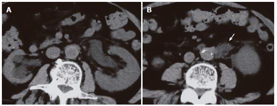
Figure 1 CT performed performed in another hospital showing left hydronephrosis (A) caused by a retroperitoneal mass (arrow) (B).
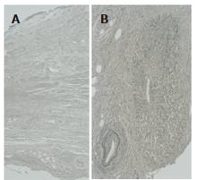
Figure 2 Histological findings of the retroperitoneal mass showing marked periureteral fibrosis with dense perivascular infiltration of lymphocytes and plasma cells (A, H&E) and obliterative phlebitis (B, Elastica Van Gienson stain).
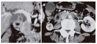
Figure 3 CT performed in our hospital showing the lower common bile duct obstructed with focally swollen pancreatic head (A) and soft tissue surrounding the aorta (arrow) (B).
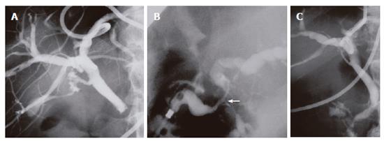
Figure 4 Percutaneous transhepatic cholangiography showing complete obstruction of the lower common bile duct (A) and resolution of the bile duct after steroid therapy (C).
Endoscopic retrograde cholangiopancreatography demonstrating segmental narrowing of the main pancreatic duct in the pancreatic head (arrow) (B).
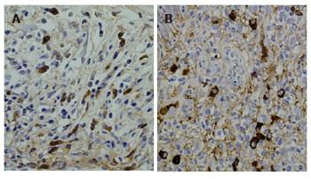
Figure 5 IgG4 immunostaining showing abundant infiltration of IgG4-positive plasma cells in tissue specimens of resected retroperitoneal mass (A) and biopsied major duodenal papilla (B).
- Citation: Kamisawa T, Chen PY, Tu Y, Nakajima H, Egawa N. Autoimmune pancreatitis metachronously associated with retroperitoneal fibrosis with IgG4-positive plasma cell infiltration. World J Gastroenterol 2006; 12(18): 2955-2957
- URL: https://www.wjgnet.com/1007-9327/full/v12/i18/2955.htm
- DOI: https://dx.doi.org/10.3748/wjg.v12.i18.2955


