Copyright
©2006 Baishideng Publishing Group Co.
World J Gastroenterol. May 14, 2006; 12(18): 2858-2863
Published online May 14, 2006. doi: 10.3748/wjg.v12.i18.2858
Published online May 14, 2006. doi: 10.3748/wjg.v12.i18.2858
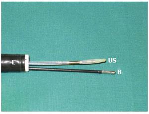
Figure 1 An endosco-pe with two channels.
A miniature ultrasound transducer (US) and a biopsy forceps (B) are inserted through the channels.
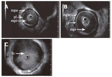
Figure 2 ES images (A and B) obtained with a 15 MHz miniprobe in a patient with achalasia showing the longitudinal, outer layer of the proper muscle (mpe) separated from a thickened circular, inner layer (mpi) by an echogenic layer corresponding to the localization of the plexus myentericus (pl).
C: shows a dilated esophageal lumen with food content (fs). A normal aspect of the proper muscle is seen.
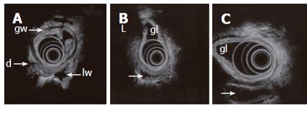
Figure 3 Endosonography with an echoendoscope (12 MHz frequency is used) placed in the gastric lumen (gl) showing the layers of the gastric wall (gw) and GI loops outside the stomach.
Lumen is partly collapsed (d), partly waterfilled (arrows). Motility and contractions of native GI loops may be studied. High frequency miniprobes are less suitable for this purpose due to higher frequencies and small transducer diameter.
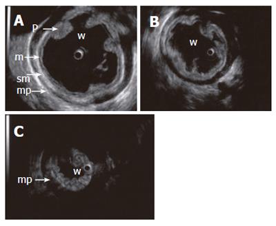
Figure 4 Contraction sequence (A, B, C) demonstrated with a 15 MHz miniprobe in a water-filled (w) stomach.
Gastric fold (p), mucosa (m), submucosa (sm), proper muscle (mp).
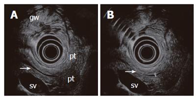
Figure 5 A normal pancreas imaged from the stomach with a 6 MHz echoendoscope.
The compressed posterior gastric wall is closely related to the surface of the pancreas. The pancreatic duct is seen (arrow) with minor (A) and slightly increased transducer pressure (B). The non-compressed gastric wall with folds (gw) is seen on the opposite side of the transducer. Pancreatic tail (pt), splenic vein (sv).
- Citation: Odegaard S, Nesje LB, Hoff DAL, Gilja OH, Gregersen H. Morphology and motor function of the gastrointestinal tract examined with endosonography. World J Gastroenterol 2006; 12(18): 2858-2863
- URL: https://www.wjgnet.com/1007-9327/full/v12/i18/2858.htm
- DOI: https://dx.doi.org/10.3748/wjg.v12.i18.2858


