Copyright
©2006 Baishideng Publishing Group Co.
World J Gastroenterol. May 7, 2006; 12(17): 2749-2755
Published online May 7, 2006. doi: 10.3748/wjg.v12.i17.2749
Published online May 7, 2006. doi: 10.3748/wjg.v12.i17.2749
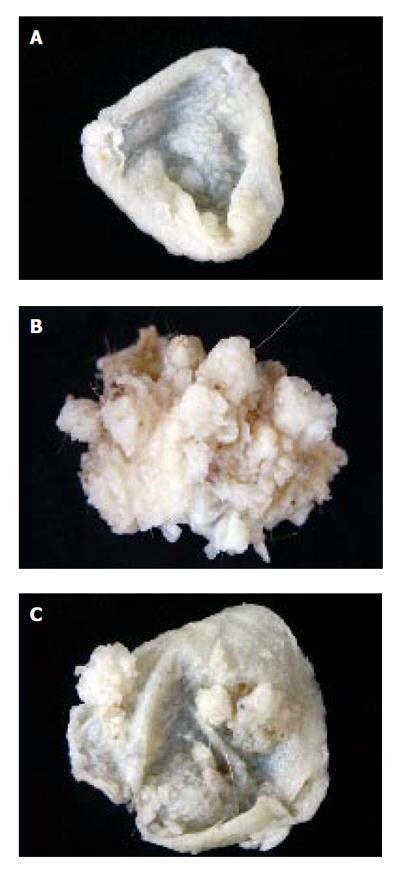
Figure 1 Gross anatomy of forestomach showing normal surface (A), tumorous surface in mice that received only B(a)P (B), and lesser number of tumors in mice that received AAILE along with B(a)P (C).
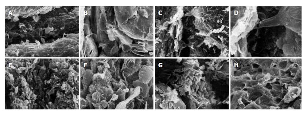
Figure 3 SEM of forestomach of mice.
A: Control mice showing grooves and ridges X 200; B: Control mice showing tongue-shaped squamous epithelium X 600; C: Mice treated with only AAILE showing grooves and ridges X 200; D: Mice treated with only AAILE showing squamous epithelial cells X 600; E: Mice received only B(a)P showing ruptured surface X 200; F: Mice received only B(a)P showing certain rounded structures in addition to tongue-shaped squamous epithelial cells X 600; G: Mice received AAILE along with B(a)P instillations showing surface rupturing X 200; H: Mice received AAILE along with B(a)P instillations showing tongue shaped squamous epithelial cells X 600.
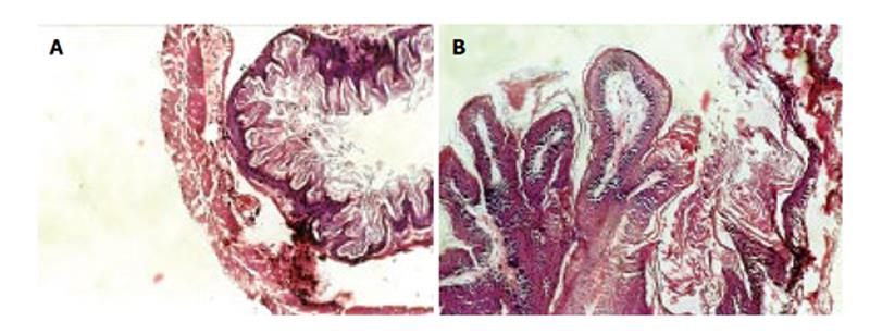
Figure 2 Histo-micrograph of forestomach.
A: Control mice showing squamous mucosa, sub-mucosa and muscular layers X 20; B: Papillary projections, disrupted submucosa and muscular layers X 20.
- Citation: Gangar SC, Sandhir R, Rai DV, Koul A. Modulatory effects of Azadirachta indica on benzo(a)pyrene-induced forestomach tumorigenesis in mice. World J Gastroenterol 2006; 12(17): 2749-2755
- URL: https://www.wjgnet.com/1007-9327/full/v12/i17/2749.htm
- DOI: https://dx.doi.org/10.3748/wjg.v12.i17.2749


