Copyright
©2006 Baishideng Publishing Group Co.
World J Gastroenterol. Apr 28, 2006; 12(16): 2625-2628
Published online Apr 28, 2006. doi: 10.3748/wjg.v12.i16.2625
Published online Apr 28, 2006. doi: 10.3748/wjg.v12.i16.2625
Figure 1 Mucosa-associated lymphoid tissue lymphoma in Patient 1.
A: A polypoid lesion on the posterior wall of the lower corpus of the stomach, suggesting the existence of lymphoproliferative disease; B: The histological features show characteristic appearance of low-grade gastric lymphomas of the mucosa-associated lymphoid tissue type with a diffuse infiltrate of centrocyte-like cells and the formation of lymphoepithelial lesions (H & E, original magnification x10).
Figure 2 Gallium scintigraphy in Patient 1 shows an abnormal accumulation of the nuclide in the para-aortic lymph nodes.
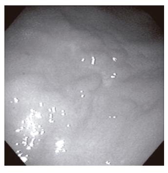
Figure 3 Twenty-four months later, an area of partly whitish rugged mucosa appeared at the site previously occupied by the mucosa-associated lymphoid tissue lymphoma in Patient 1.
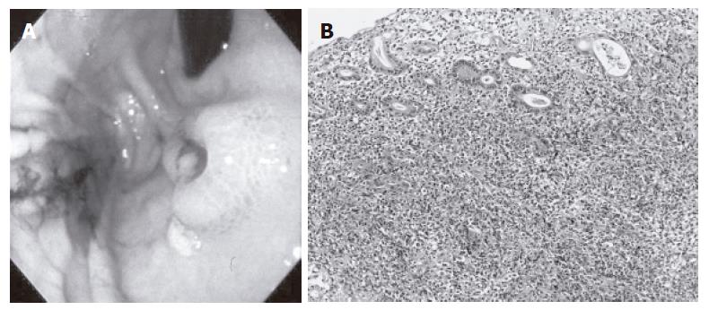
Figure 4 Mucosa-associated lymphoid tissue lymphoma in Patient 2.
A: An ulcerated lesion on the anterior wall of the upper corpus before eradication; B: The histological features show the appearance of low-grade gastric lymphomas of the mucosa-associated lymphoid tissue type similar to that found in Patient 1 (H & E, original magnification x10).
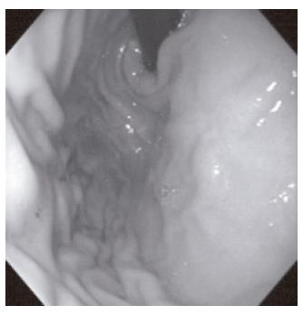
Figure 5 Two months after eradication therapy in Patient 2, an area of glossy whitish mucosa appeared at the site previously occupied by the low-grade gastric lymphoma of the mucosa-associated lymphoid tissue type.
- Citation: Ohno Y, Kosaka T, Muraoka I, Kanematsu T, Tsuru A, Kinoshita E, Moriuchi H. Remission of primary low-grade gastric lymphomas of the mucosa-associated lymphoid tissue type in immunocompromised pediatric patients. World J Gastroenterol 2006; 12(16): 2625-2628
- URL: https://www.wjgnet.com/1007-9327/full/v12/i16/2625.htm
- DOI: https://dx.doi.org/10.3748/wjg.v12.i16.2625


