Copyright
©2006 Baishideng Publishing Group Co.
World J Gastroenterol. Apr 28, 2006; 12(16): 2549-2555
Published online Apr 28, 2006. doi: 10.3748/wjg.v12.i16.2549
Published online Apr 28, 2006. doi: 10.3748/wjg.v12.i16.2549
Figure 1 Immunohistochemical staining of lymph node micrometastasis with the monoclonal antibody CAM 5.
2. A: Micrometastasis consisting of a single cell (original magnification, × 200). B: Micrometastasis consisting of a small cluster of tumor cells (original magnification, × 100).
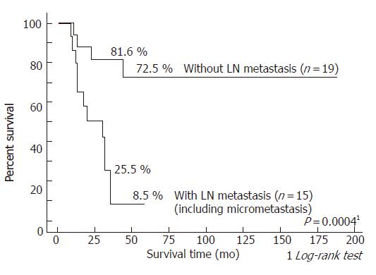
Figure 2 Survival curves after resection for hilar bile duct carcinoma according to the presence of lymph node metastasis, including micrometastasis.
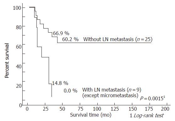
Figure 3 Survival curves after resection for hilar bile duct carcinoma according to the presence of lymph node metastasis: patients without lymph node metastasis versus those with overt lymph node and micro metastasis.
Figure 4 Survival curves after resection for hilar bile duct carcinoma according to the presence of lymph node metastasis: patients without lymph node metastasis and those with lymph node micrometastasis versus those with overt lymph node metastasis.
Figure 5 Immunohistochemical staining of primary tumors with VEGF-C polyclonal antibody.
A: VEGF-C positive (original magnification, × 400). B: VEGF-C-negative (original magnification, × 200).
- Citation: Taniguchi K, Iida T, Hori T, Yagi S, Imai H, Shiraishi T, Uemoto S. Impact of lymph node micrometastasis in hilar bile duct carcinoma patients. World J Gastroenterol 2006; 12(16): 2549-2555
- URL: https://www.wjgnet.com/1007-9327/full/v12/i16/2549.htm
- DOI: https://dx.doi.org/10.3748/wjg.v12.i16.2549


