Copyright
©2006 Baishideng Publishing Group Co.
World J Gastroenterol. Apr 21, 2006; 12(15): 2464-2465
Published online Apr 21, 2006. doi: 10.3748/wjg.v12.i15.2464
Published online Apr 21, 2006. doi: 10.3748/wjg.v12.i15.2464
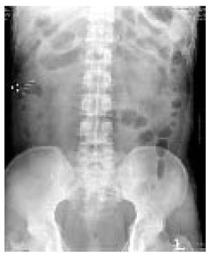
Figure 1 A plain abdominal radiograph showing a characteristic toothbrush image with parallel rows of short metallic radiodensities in the right upper quadrant (arrow).
Figure 2 Abdominal computed tomography (CT) imaging.
A: A metallic density in the ascending colon (arrow); B: A low density lesion penetrating the lateral section of the liver (arrow).
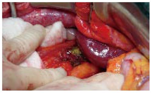
Figure 3 Shaft of a toothbrush penetrating the proximal transverse colon and the lateral section of the liver (arrow).
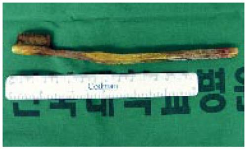
Figure 4 Extracted 20 cm toothbrush.
- Citation: Lee MR, Hwang Y, Kim JH. A case of colohepatic penetration by a swallowed toothbrush. World J Gastroenterol 2006; 12(15): 2464-2465
- URL: https://www.wjgnet.com/1007-9327/full/v12/i15/2464.htm
- DOI: https://dx.doi.org/10.3748/wjg.v12.i15.2464


