Copyright
©2006 Baishideng Publishing Group Co.
World J Gastroenterol. Apr 21, 2006; 12(15): 2394-2397
Published online Apr 21, 2006. doi: 10.3748/wjg.v12.i15.2394
Published online Apr 21, 2006. doi: 10.3748/wjg.v12.i15.2394
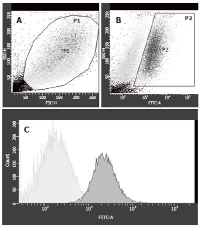
Figure 1 FACS sorting of GFP+ and GFP- cells of co-cultures at wk 1.
A: Viable cells were gated as P1 cells; B: P1 cells were gated in GFP- or GFP+ (P2); C: For the highest purification of GFP+ cells, P2 cells were analyzed and only GFP+ cells were sorted for PCR-analysis (light grey peak), 34.5 % of cells from P2 were GFP+ and 65.5 % were GFP-.
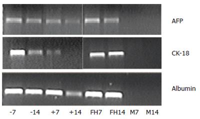
Figure 2 Expressions of the liver specific markers (AFP, CK-18, or albumin) of cultured rMSCs (M), fetal liver cells (FH), and GFP+ (+) or GFP- (-) cells of the co-cultures on d 7 or 14.
Cultured rMSCs lacked expression of AFP, CK-18, or albumin on d 7 or 14 (lanes M7 and M14). Cultured fetal liver cells showed expression of AFP, CK-18 and albumin on d 7 or 14 (lanes FH7 and FH14). The GFP+ cells (lanes +7 and +14) showed clear expression of albumin and AFP, and lower expression of CK-18. The GFP- cells (lanes -7 and -14) showed a stable expression of AFP, CK-18, and albumin.
- Citation: Lange C, Bruns H, Kluth D, Zander AR, Fiegel HC. Hepatocytic differentiation of mesenchymal stem cells in cocultures with fetal liver cells. World J Gastroenterol 2006; 12(15): 2394-2397
- URL: https://www.wjgnet.com/1007-9327/full/v12/i15/2394.htm
- DOI: https://dx.doi.org/10.3748/wjg.v12.i15.2394


