Copyright
©2006 Baishideng Publishing Group Co.
World J Gastroenterol. Apr 14, 2006; 12(14): 2209-2216
Published online Apr 14, 2006. doi: 10.3748/wjg.v12.i14.2209
Published online Apr 14, 2006. doi: 10.3748/wjg.v12.i14.2209
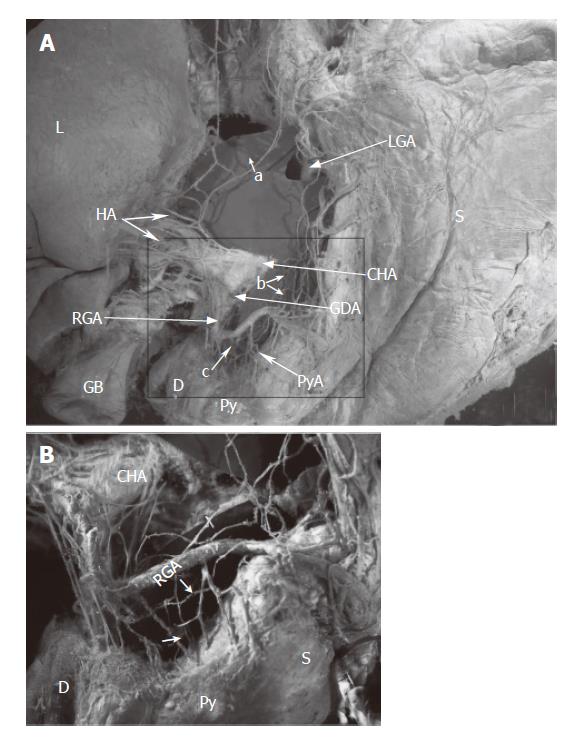
Figure 1 The innervation of the pyloric region from the view of the superior part of the pylorus, its schematic shows in Figure 3A.
Hepatic divisions (a) join in the anterior hepatic plexus in the proper hepatic artery or the left/right hepatic artery (HA). The nerves ran along the right gastric artery (RGA) and its branches (PyA), and reached the pyloric region (Py) (c). The nerves (b) of Latarjet ran along the lesser curvature or the branches of the left gastric artery (LGA), intended for the antro-pyloric region. B is an enlargement of the box in A., the arrows show the nerves innervating the pyloric region from the right gastric artery. CHA, common hepatic artery; D, duodenum; E, esophagus; GB, gallbladder; GDA, gastroduodenal artery; L, liver; S, stomach.
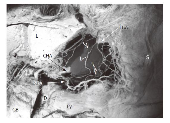
Figure 2 Another specimen, its schematic shows in Figure 3B, showing a descending branch (b) originating from the hepatic division of the anterior vagal trunk passing through the hepatogastric ligament, obliquely downward, reaching the right gastric artery (RGA), innervating the pyloric region (Py).
a is similar to a in Figure 1A, indicating the hepatic divisions. c, indicating the branches for the pyloric region from the nerves of Latarjet. CHA, common hepatic artery; D, duodenum; GB, gallbladder; L, liver; LGA, left gastric artery; S, stomach.
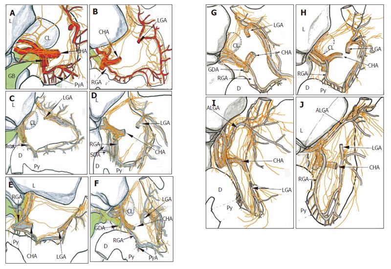
Figure 3 Diagram indicating the distribution in and around the cardia, the lesser curvature, the porta hepatica and the antro-pyloric region in 10 cadavers.
Among them, A and B are diagrams of Figures 1A and 2, respectively. Five specimens, B, D, F, I and J, showed hepatic divisions joining directly to the right gastric artery, while, for the other specimens, after joining to the proper hepatic or hepatic artery, the nerves sent off some offshoots to the right gastric artery, and innervated the pyloric region. ALGA, accessory left gastric artery; CHA, common hepatic artery; CL, caudal liver; GB, gallbladder; L, liver; LGA, left gastric artery; Py, pylorus; PyA, pyloric artery; RGA, right gastric artery; SDA, supra-duodenal artery.
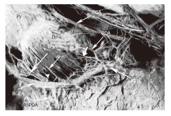
Figure 4 An example of innervation in the posterior part of the pylorus.
The stomach was raised. The nerves originating from the right gastroepiploic artery (RGEA) or the gastroduodenal artery (GDA) running along the retroduodenal artery (RDA) (white arrow) or the infrapyloric artery (IPyA) (white arrowhead) reached the first duodenum posterior part and the pylorus of the posterior part.
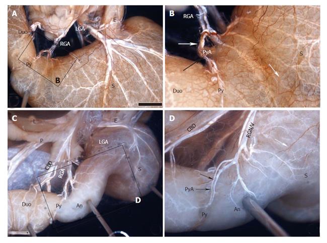
Figure 5 Two cases showing innervation of the pyloric region (Py) in Suncus murinus by whole-mount immunostaining.
High magnifications of the boxed areas in A and C are shown in B and D. White arrows show the nerves of Latarjet intended for the antro-pyloric region. Black arrows indicate the nerves arising from the right gastric artery (RGA), running along the pyloric artery (PyA), and reaching the pyloric region. An, pyloric antrum; CBD, common bile duct; Duo, duodenum; E, esophagus; L, liver; LGA, left gastric artery; P, pancreas; RGA/V, right gastric artery/vein; S, stomach. Scale bar = 2 mm in A and B.
- Citation: Yi SQ, Ru F, Ohta T, Terayama H, Naito M, Hayashi S, Buhe S, Yi N, Miyaki T, Tanaka S, Itoh M. Surgical anatomy of the innervation of pylorus in human and Suncus murinus, in relation to surgical technique for pylorus-preserving pancreaticoduodenectomy. World J Gastroenterol 2006; 12(14): 2209-2216
- URL: https://www.wjgnet.com/1007-9327/full/v12/i14/2209.htm
- DOI: https://dx.doi.org/10.3748/wjg.v12.i14.2209


