Copyright
©2006 Baishideng Publishing Group Co.
World J Gastroenterol. Mar 28, 2006; 12(12): 1975-1977
Published online Mar 28, 2006. doi: 10.3748/wjg.v12.i12.1975
Published online Mar 28, 2006. doi: 10.3748/wjg.v12.i12.1975
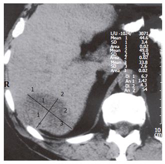
Figure 1 Abdominal CT showing presence of the right lobe tumor of the liver.
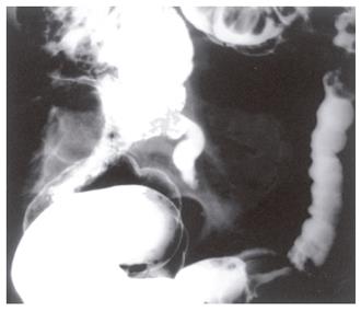
Figure 2 Barium enema displaying extraluminal compression and medial dislocation of the cecum due to cystadenoma of the appendix and tubular stenosis of the sigmoid colon due to the adenocarcinoma.
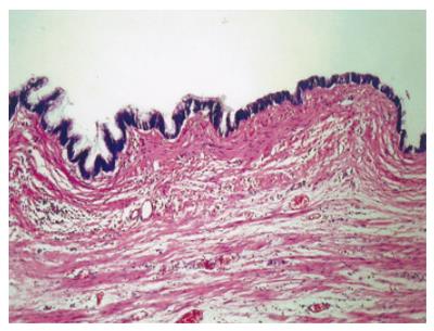
Figure 3 Cystadenoma mucinosum appendicis with obvious dysplastic epithelial lining and focally evident mucinous cytoplasmatic production (H&E, 64x).
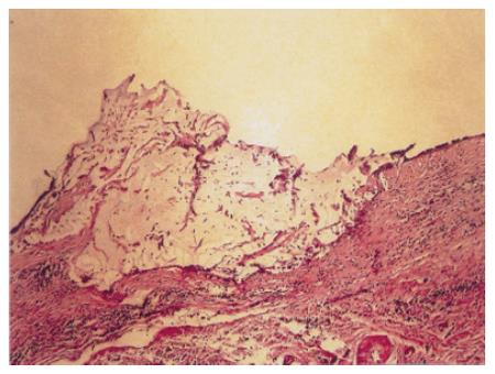
Figure 4 Cystadenoma mucinosum appendices with small extracellular mucinous deposit just beneath focally eroded adenomatous epithelium (H&E, 13x).
- Citation: Djuranovic SP, Spuran MM, Kovacevic NV, Ugljesic MB, Kecmanovic DM, Micev MT. Mucinous cystadenoma of the appendix associated with adenocarcinoma of the sigmoid colon and hepatocellular carcinoma of the liver: Report of a case. World J Gastroenterol 2006; 12(12): 1975-1977
- URL: https://www.wjgnet.com/1007-9327/full/v12/i12/1975.htm
- DOI: https://dx.doi.org/10.3748/wjg.v12.i12.1975


