Copyright
©2006 Baishideng Publishing Group Co.
World J Gastroenterol. Mar 14, 2006; 12(10): 1603-1606
Published online Mar 14, 2006. doi: 10.3748/wjg.v12.i10.1603
Published online Mar 14, 2006. doi: 10.3748/wjg.v12.i10.1603
Figure 1 T1-weighted axial SE contrast-unenhanced (A) and MnDPDP-enhanced (B) images of pancreatic adenocarcinoma with liver metastases.
MnDPDP enhancement was not determined in the lesion while the normal parenchyma of the pancreas showed homogeneous contrast enhancement. MnDPDP- enhanced image showed the better delination of the tumor (thick arrows). Tiny liver metastases (thin arrows) were not determined on unenhanced MR image but on MnDPDP-enhanced MRI.
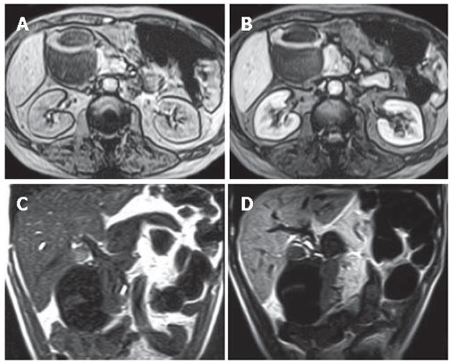
Figure 2 Unenhanced (A) and MnDPDP-enhanced (B) axial T1-weighted GRE images, unenhanced (C) and MnDPDP-enhanced (D) coronal T1-weighted SE images in pancreatic parenchyma out of lesion and liver parenchyma after MnDPDP infusion.
Minimal contrast enhancement in the septa of mass was found on MnDPDP-enhanced images (B and D).
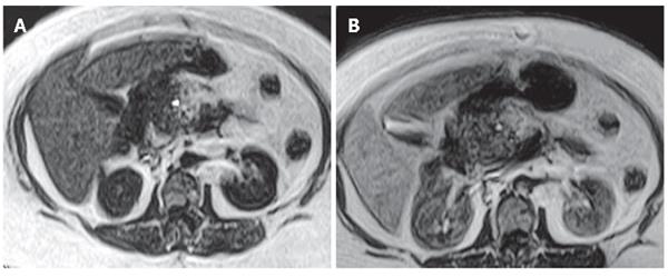
Figure 3 Contrast-unenhanced T1-weighted axial SE image demonstrating focal enlargement of the pancreatic head with a low signal intensity, simulating the appearence of pancreatic cancer (A) and MnDPDP-enhanced T1-weighted axial SE image showing heterogeneous and reduced contrast enhancement in this focal lesion (B).
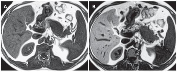
Figure 4 Unenhanced (A) and MnDPDP-enhanced (B) axial T1-weighted SE images showing better delineation of the tumor after MnDPDP infusion.
- Citation: Eser G, Karabacakoglu A, Karakose S, Eser C, Kayacetin E. Mangafodipir trisodium-enhanced magnetic resonance imaging for evaluation of pancreatic mass and mass-like lesions. World J Gastroenterol 2006; 12(10): 1603-1606
- URL: https://www.wjgnet.com/1007-9327/full/v12/i10/1603.htm
- DOI: https://dx.doi.org/10.3748/wjg.v12.i10.1603


