Copyright
©2006 Baishideng Publishing Group Co.
World J Gastroenterol. Mar 14, 2006; 12(10): 1529-1535
Published online Mar 14, 2006. doi: 10.3748/wjg.v12.i10.1529
Published online Mar 14, 2006. doi: 10.3748/wjg.v12.i10.1529
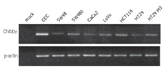
Figure 1 PCR amplification of the complete coding region of Chibby mRNA.
The Chibby coding region was amplified using RT-PCR. All colon carcinoma cell lines were shown to express full-length Chibby mRNA. Expression levels cannot be estimated from this experiment as the PCR was carried out to saturation in order to illustrate full-length mRNA expression and to obtain enough PCR products for sequencing. β-actin amplification served as control to ascertain the integrity of the cDNA in all samples.
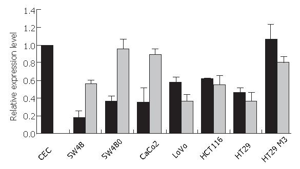
Figure 2 Quantification of Chibby expression in colon carcinoma cell lines.
The amount of Chibby mRNA expression was carefully quantified by real-time PCR. All colon carcinoma cell lines showed down-regulation of Chibby mRNA expression in comparison to colon epithelial cells (CEC) and HT29 M3 cells, a cell line derived from HT29 cells that is considered to be a highly differentiated colon carcinoma cell line (black bars). Treatment of cells with the demethylating agent 5-azacytidine caused a modest increase in Chibby mRNA levels in SW48, SW480, and CaCo2 cells, but not in the other cell lines (grey bars). HT29 M3 cells were used as controls for the demethylation experiments as the experiment could not have been performed with primary CEC due to culture conditions.
Figure 3 Expression of Chibby in transfected SW480 cells.
A: Western blot analysis showing Chibby protein expression levels in untransfected (mock), control vector, or Chibby transfected SW480 cells. B: immunofluorescence staining of flag-tagged Chibby transfected SW480 cells. Nuclei were stained with DAPI in blue, flag-tagged Chibby was detected with FITC-labelled anti-flag antibody (green). C: TOPflash reporter gene assays showing inhibition of β-catenin signalling after transfection of SW480 cells with Chibby expression plasmid.
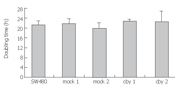
Figure 4 Proliferation Assay.
Analysis of the cells over-expressing Chibby could not altered doubling time in comparison to mock or control transfected SW480 cells. Assays were performed in triplicates.
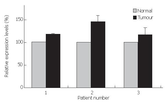
Figure 5 Chibby mRNA expression levels in colorectal tumor and corresponding normal tissue samples.
Quantitative RT-PCR showing Chibby mRNA expression levels in matched pairs of tumor and normal colorectal tissue of 3 different patients.
- Citation: Schuierer MM, Graf E, Takemaru KI, Dietmaier W, Bosserhoff AK. Reduced expression of β-catenin inhibitor Chibby in colon carcinoma cell lines. World J Gastroenterol 2006; 12(10): 1529-1535
- URL: https://www.wjgnet.com/1007-9327/full/v12/i10/1529.htm
- DOI: https://dx.doi.org/10.3748/wjg.v12.i10.1529


