Copyright
©2005 Baishideng Publishing Group Inc.
World J Gastroenterol. Feb 28, 2005; 11(8): 1131-1140
Published online Feb 28, 2005. doi: 10.3748/wjg.v11.i8.1131
Published online Feb 28, 2005. doi: 10.3748/wjg.v11.i8.1131
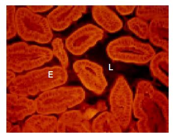
Figure 1 I = ileum of a wild type normal mouse narrow and free of bacteria in most parts.
E = epithelium; L = lumen.
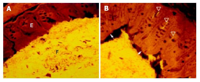
Figure 2 Concentrated bacterial mass in direct contact with the mucosal surface (A) and non-adherent bacteria (B) in cecum of a healthy wild type mouse.
E = epithelium; F = feces. Arrow indicates the shrinkage of feces, arrowheads indicate bacteria in crypts.
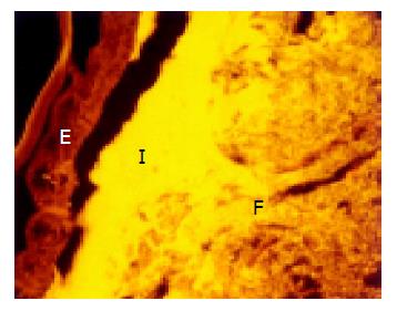
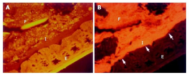
Figure 3 Interlaced layer in the distal portion of the proximal colon.
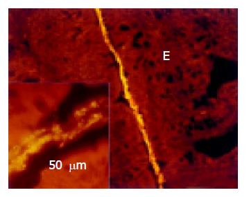
Figure 4 Middle colon in a normal mouse.
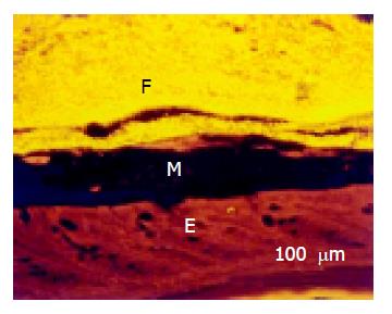
Figure 5 Rectal mucosa covered with thick mucus (M).
E = epithelium, F = feces.
Figure 6 Fecal bacteria hybridized with Bacteroides (Cy3 green-orange, 6A) and Erec (Cy5 red, 6B) probes, cecum of healthy WT mice.
Figure 7 Interlaced layer in the proximal colon of IL-10 mice with colitis visualized simultaneously by hybridization with Bac303 (Cy3 green-orange, 7A) and Eub338 probes (Cy5 red, 7B).
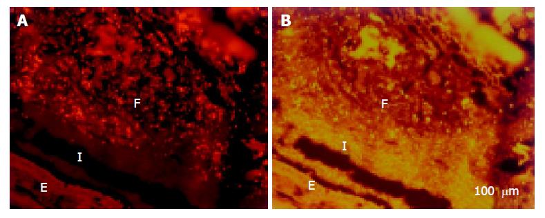
Figure 8 Interlaced layer in proximal colon of DSS mice visualized simultaneously by hybridization with Clit135 (Cy5 red, 8A) and Lab158 probes (Cy3 green-orange, 8B).
I = interlaced layer, E = epithelium, F = feces.
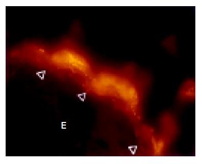
Figure 9 Bacteria location below the intact mucus layer (arrowheads) and adherence to the colonic mucosa in DSS-exposed mice.
Figure 10 Bacteria infiltrating the mucosa in distal colon of IL-10 mice with colitis (arrowheads).
-
Citation: Swidsinski A, Loening-Baucke V, Lochs H, Hale LP. Spatial organization of bacterial flora in normal and inflamed intestine: A fluorescence
in situ hybridization study in mice. World J Gastroenterol 2005; 11(8): 1131-1140 - URL: https://www.wjgnet.com/1007-9327/full/v11/i8/1131.htm
- DOI: https://dx.doi.org/10.3748/wjg.v11.i8.1131


