Copyright
©2005 Baishideng Publishing Group Inc.
World J Gastroenterol. Feb 7, 2005; 11(5): 767-769
Published online Feb 7, 2005. doi: 10.3748/wjg.v11.i5.767
Published online Feb 7, 2005. doi: 10.3748/wjg.v11.i5.767
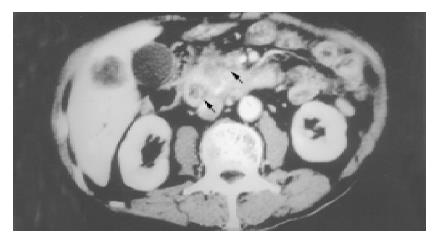
Figure 1 A soft tissue mass (3 cm×3 cm in size) over the pancreatic head (arrow), duodenal infiltration (arrowhead), and superior mesenteric vein encasement shown by abdominal computed tomography.
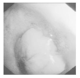
Figure 2 A soft polypoid mass protruding over the anterior wall of the duodenal bulb with partial obstruction of the second portion of the duodenum shown by upper gastrointestinal endoscopy.
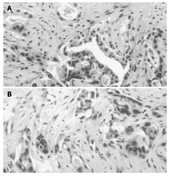
Figure 3 Morphology of adenocarcinoma and pancreatic lesion.
A: Biopsy of the duodenal tumor via upper gastrointestinal endoscopy revealed a picture of adenocarcinoma cells in an acinar pattern; B: Biopsy of the pancreatic head tumor via computed tomography-guidance showed moderately differentiated adenocarcinoma cells infiltrating into the collagenous stroma (hematoxylin and eosin stain; magnification ×200).
- Citation: Lin YH, Chen CY, Chen CP, Kuo TY, Chang FY, Lee SD. Hematemesis as the initial complication of pancreatic adenocarcinoma directly invading the duodenum: A case report. World J Gastroenterol 2005; 11(5): 767-769
- URL: https://www.wjgnet.com/1007-9327/full/v11/i5/767.htm
- DOI: https://dx.doi.org/10.3748/wjg.v11.i5.767


