Copyright
©2005 Baishideng Publishing Group Inc.
World J Gastroenterol. Feb 7, 2005; 11(5): 629-633
Published online Feb 7, 2005. doi: 10.3748/wjg.v11.i5.629
Published online Feb 7, 2005. doi: 10.3748/wjg.v11.i5.629
Figure 1 In situ hybridization for EBER1 in gastric carcinoma tissue.
A: In situ EBER1 hybridization with antisense probes. Strong signals were observed in the nuclei of all tumor cells; B: In situ EBER1 hybridization with sense probes; C: Hematoxylin/eosin (H&E) staining in the adjacent pair of EBER1 in situ hybridization. original magnification ×200.
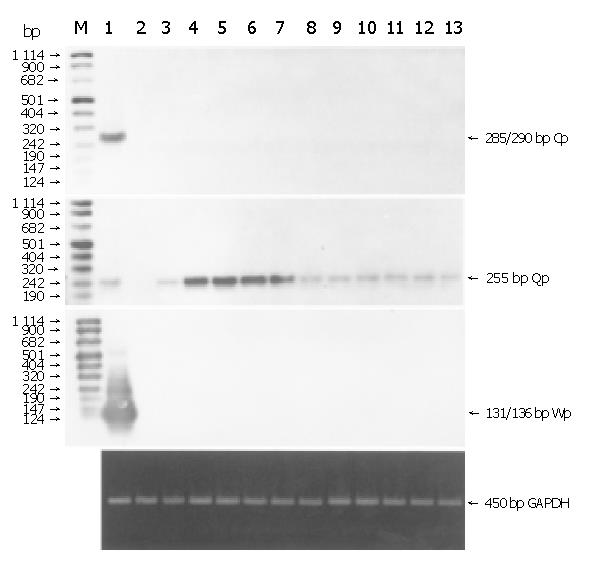
Figure 2 RT-PCR and Southern hybridization analysis of EBNA gene transcription using promoters.
M: DIG-labeled DNA molecular weight marker VIII (Roche). Lane 1: EBV-positive LCL(positive control); lane 2: EBV-negative Ramos cells(negative control); lanes 3-13: EBV-positive gastric carcinoma samples. GAPDH mRNA was amplified to check pertinent RNA extraction and results were shown by EB staining.
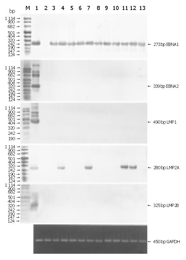
Figure 3 Detection of EBV latent gene expression (A) and EBV lytic gene expression (B) by RT-PCR and Southern hybridization.
M: DIG-labeled DNA molecular weight marker VIII (Roche). Lane 1: EBV-positive LCL (positive control); lane 2: EBV-negative Ramos cells (negative control); lanes 3-13: EBV-positive gastric carcinoma samples. GAPDH mRNA was amplified to check pertinent RNA extraction and results were shown by EB staining.
- Citation: Luo B, Wang Y, Wang XF, Liang H, Yan LP, Huang BH, Zhao P. Expression of Epstein-Barr virus genes in EBV-associated gastric carcinomas. World J Gastroenterol 2005; 11(5): 629-633
- URL: https://www.wjgnet.com/1007-9327/full/v11/i5/629.htm
- DOI: https://dx.doi.org/10.3748/wjg.v11.i5.629


