Copyright
©2005 Baishideng Publishing Group Inc.
World J Gastroenterol. Dec 28, 2005; 11(48): 7585-7590
Published online Dec 28, 2005. doi: 10.3748/wjg.v11.i48.7585
Published online Dec 28, 2005. doi: 10.3748/wjg.v11.i48.7585
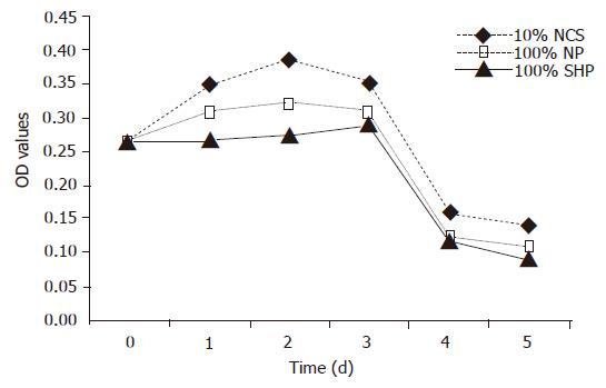
Figure 1 Viability of porcine hepatocytes cultured in ♦ medium containing 10% NCS, □ 100% NP, ▲ 100% SVHP.
Results were expressed as mean±SD for six samples.
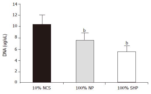
Figure 2 DNA content in porcine hepatocytes grown in three different media.
DNA synthesis in 100% SVHP and normal plasma was compared to that in the medium containing 10% NCS (bP<0.01 vs 10% NCS group, by ANOVA followed by multiple comparisons). Results were expressed as mean±SD for six samples Black oblique line: 10% NCS, black small point: 100% NP; black small square: 100% SVHP.
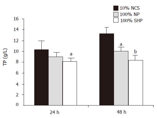
Figure 3 TP content in porcine hepatocytes grown in three different media.
TP synthesis in 100% SVHP and NP was compared to that in the medium containing 10% NCS (aP<0.05, bP<0.01 vs 10% NCS group, by two-way variance analysis). Results were expressed as mean±SD for six samples. Black bar: 10% NCS; gray bar: 100% NP; white bar: 100% SVHP.
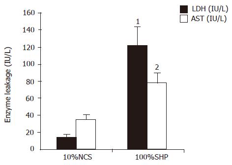
Figure 4 Leakage of LDH and AST after 5 h of culture (black bar: LDH; white bar: AST).
The levels of LDH and AST in 100% SVHP were significantly higher than those in the medium containing 10% NCS. Results were expressed as mean±SD (1: t = 24.552, P = 0.001 and 2: t = 4.169, P = 0.014, compared to the10% NCS group, n = 6).
Figure 5 GSH content in porcine hepatocytes grown in three different cultures (A) (aP<0.
05 vs 10% NCS, by two-way variance analysis, compared to 10% NCS) and CAT activity of porcine hepatocytes grown in the three different cultures (B) (cP<0.05 vs 10% NCS by two-way variance analysis, compared to 10% NCS). Black bar: 10% NCS, gray bar: 100% NP; white bar: 100% SVHP.
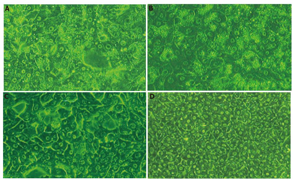
Figure 6 Morphological changes of porcine hepatocytes cultured in 100% SVHP (A and B) and media containing 10% NCS (C and D).
After 24 h of culture, hepatocytes in the100% SVHP group were detached from the dishes, vacuolization in the cytoplasm and deformities were more commonly observed. The membranes of cells became indistinct (A); after 48 h of culture, vacuoles were more and mainly concentrated around cell nuclei (B); after 24 h of culture, hepatocytes in the 10% NCS group constituted confluent monolayers with an intact morphology throughout the culture (C); after 48 h of culture, hepatocytes in the 10% NCS group constituted confluent monolayers with an intact morphology throughout the culture (D).
- Citation: Cheng YB, Wang YJ, Zhang SC, Liu J, Chen Z, Li JJ. Response of porcine hepatocytes in primary culture to plasma from severe viral hepatitis patients. World J Gastroenterol 2005; 11(48): 7585-7590
- URL: https://www.wjgnet.com/1007-9327/full/v11/i48/7585.htm
- DOI: https://dx.doi.org/10.3748/wjg.v11.i48.7585


