Copyright
©2005 Baishideng Publishing Group Inc.
World J Gastroenterol. Dec 21, 2005; 11(47): 7531-7535
Published online Dec 21, 2005. doi: 10.3748/wjg.v11.i47.7531
Published online Dec 21, 2005. doi: 10.3748/wjg.v11.i47.7531
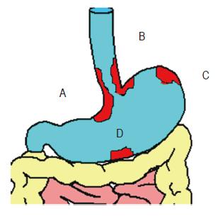
Figure 1 Classification of GVs on the basis of location and relationship with EVs (A and B: GOV, gastroesophageal varices, type 1 and 2 respectively.
C and D: IGV, isolated GVs, type 1 and 2 respectively).
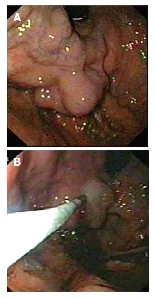
Figure 2 (A) (top) GVs at fundus, (B) (bottom) cyanoacrylate injection into the GV.
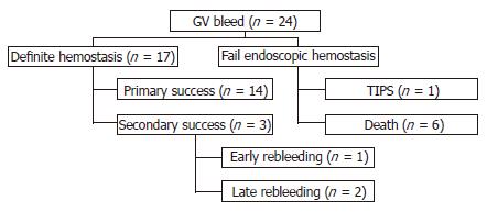
Figure 3 Diagram demonstrating efficacy of cyanoacrylate glue for the treatment of GV bleeding.
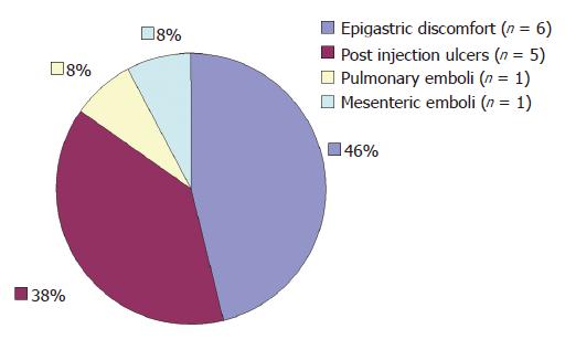
Figure 4 Complications of cyanoacrylate glue injection.
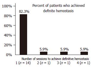
Figure 5 Number of sessions and percent of patients who achieved definite hemostasis (n = 17).
- Citation: Noophun P, Kongkam P, Gonlachanvit S, Rerknimitr R. Bleeding gastric varices: Results of endoscopic injection with cyanoacrylate at King Chulalongkorn Memorial Hospital. World J Gastroenterol 2005; 11(47): 7531-7535
- URL: https://www.wjgnet.com/1007-9327/full/v11/i47/7531.htm
- DOI: https://dx.doi.org/10.3748/wjg.v11.i47.7531


