Copyright
©2005 Baishideng Publishing Group Inc.
World J Gastroenterol. Dec 14, 2005; 11(46): 7359-7363
Published online Dec 14, 2005. doi: 10.3748/wjg.v11.i46.7359
Published online Dec 14, 2005. doi: 10.3748/wjg.v11.i46.7359
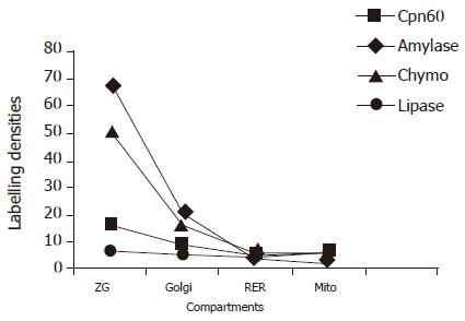
Figure 1 Distribution of Cpn60 and pancreatic enzymes in the compartments of the pancreatic acinar cells in normal rats.
Labeling densities (gold particles/μm2) were measured by immunocytochemistry. ZG: zymogen granules; RER: rough endoplasmic reticulum; Mito: mitochondria; Chymo: chymotrypsinogen.
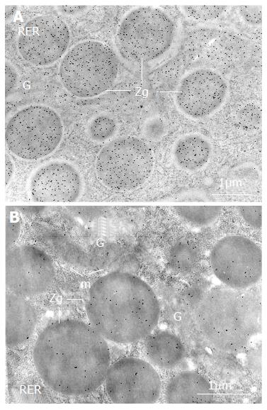
Figure 2 Changes of Cpn60 and pancreatic enzymes in the acinar cells of diabetic rats.
The labeling by gold particles (black particles), reflecting Cpn60 antigenic sites, is more intense over the zymogen granules (ZG) in the pancreatic acinar cell in diabetic rat (A) compared with that in normal rat (B). RER: rough endoplasmic reticulum; G: Golgi; m: mitochondria.
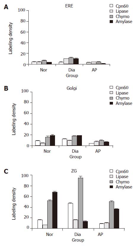
Figure 3 Immunolabeling densities of Cpn60 and the pancreatic enzymes in pancreatic acinar cells rats (particles/μm2, mean± SE).
A: Rough endoplasmic reticulum; B: Golgi; and C: zymogen granules. Nor: normal group; Dia: diabetic group; Chymo: chymotrypsinogen.
- Citation: Li YY, Bendayan M. Alteration of chaperonin60 and pancreatic enzyme in pancreatic acinar cell under pathological condition. World J Gastroenterol 2005; 11(46): 7359-7363
- URL: https://www.wjgnet.com/1007-9327/full/v11/i46/7359.htm
- DOI: https://dx.doi.org/10.3748/wjg.v11.i46.7359


