Copyright
©2005 Baishideng Publishing Group Inc.
World J Gastroenterol. Dec 14, 2005; 11(46): 7254-7260
Published online Dec 14, 2005. doi: 10.3748/wjg.v11.i46.7254
Published online Dec 14, 2005. doi: 10.3748/wjg.v11.i46.7254
Figure 1 Changes in vascular endothelial growth factor (VEGF), angiopoietin (Ang) and Tie-2 mRNA expressions after 70% hepatectomy.
A: Quantification of VEGF mRNA levels after hepatectomy by RT-PCR using a LightCycler (Roche Diagnostics). aP<0.05 vs 0 h; B: quantification of Ang-1 (closed circle), Ang-2 (open circle), and Tie-2 (open bar) mRNA levels after hepatectomy by RT-PCR using a LightCycler. aP<0.05 vs 0 h, 1P<0.03 vs 0 h, respectively.
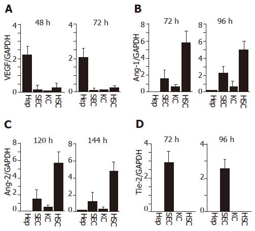
Figure 2 Expressions of VEGF (A), Ang-1 (B), Ang-2 (C) and Tie-2 mRNA (D) in isolated liver cells at different time points.
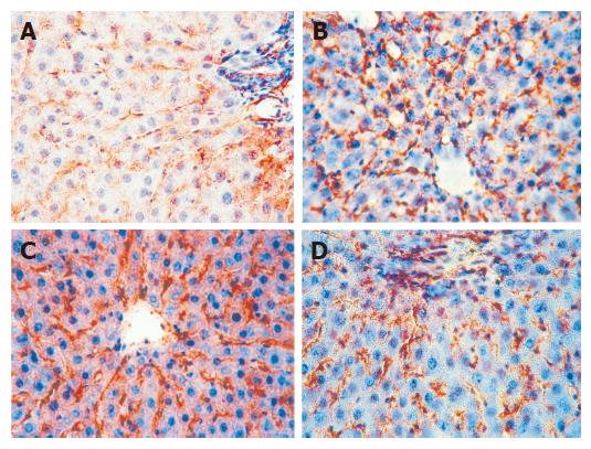
Figure 3 Immunohistochemical staining for angiopoietins (Ang) and Tie-2 protein.
Ang-1 protein before hepatectomy (A), 96 h after hepatectomy (B), Ang-2 protein induction 120 h after hepatectomy (C), Tie-2 protein before hepatectomy (D) (original magnification x 200).
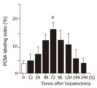
Figure 4 PCNA labeling index of sinusoidal endothelial cells (SECs) after 70 % hepatectomy in rats.
aP<0.05 vs 0 h.
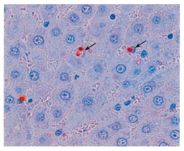
Figure 5 TUNEL staining of regenerating liver tissue at 120 h after hepatectomy.
Arrows indicate TUNEL stained-positive sinusoidal cells.
- Citation: Shimizu H, Mitsuhashi N, Ohtsuka M, Ito H, Kimura F, Ambiru S, Togawa A, Yoshidome H, Kato A, Miyazaki M. Vascular endothelial growth factor and angiopoietins regulate sinusoidal regeneration and remodeling after partial hepatectomy in rats. World J Gastroenterol 2005; 11(46): 7254-7260
- URL: https://www.wjgnet.com/1007-9327/full/v11/i46/7254.htm
- DOI: https://dx.doi.org/10.3748/wjg.v11.i46.7254


