Copyright
©2005 Baishideng Publishing Group Inc.
World J Gastroenterol. Dec 7, 2005; 11(45): 7218-7221
Published online Dec 7, 2005. doi: 10.3748/wjg.v11.i45.7218
Published online Dec 7, 2005. doi: 10.3748/wjg.v11.i45.7218
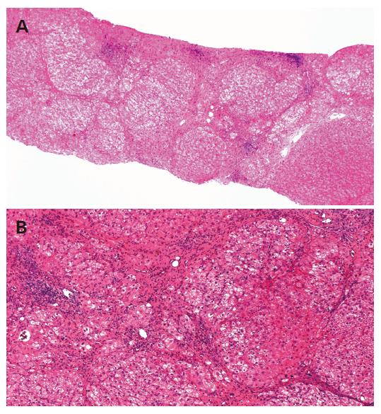
Figure 1 (A and B) Liver biopsy specimen before interferon therapy revealed liver cirrhosis with mild inflammation.
It was graded as A1F4 according to the Classification by Desmet. Alcohol hyaline was not found (H&E stain, original magnifications A: ×4, B: ×200).
Figure 2 Contrast medium-enhanced CT image of the liver tumor of the right inferior segment (a white arrow).
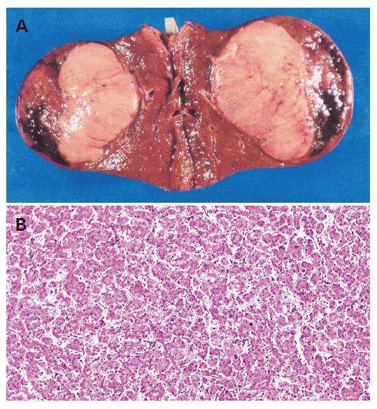
Figure 3 (A) Surgical specimen showing an 8-cm tumor.
(B) Histological examination revealing a well-differentiated hepatocellular carcinoma (H&E stain, original magnification ×200).
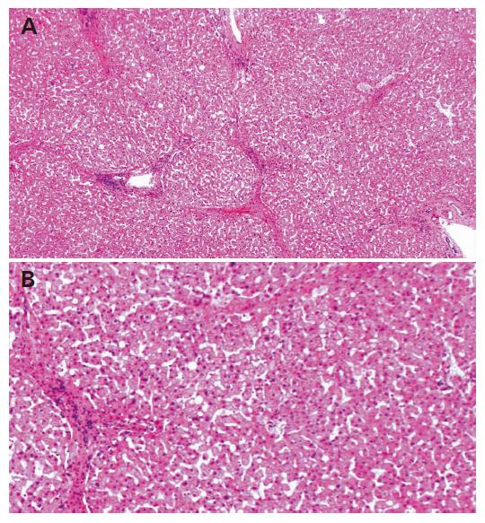
Figure 4 (A and B) Surgical specimen of non-cancerous lesion showing improvement of fibrosis and inflammation.
However, great amounts of septa were observed, although fibrous bundles apparently decreased. Fibrosis resolved, revealing liver cirrhosis with mild inflammation. It was graded as A1F3 (H&E stain, original magnifications A: ×4, B: ×200).
- Citation: Ito Y, Yamamoto N, Nakata R, Kato Y, Iori M, Sakai K, Takemura T, Tateishi R, Yoshida H, Kawabe T, Omata M. Delayed development of hepatocellular carcinoma during long-term follow-up after eradication of hepatitis C virus by interferon therapy. World J Gastroenterol 2005; 11(45): 7218-7221
- URL: https://www.wjgnet.com/1007-9327/full/v11/i45/7218.htm
- DOI: https://dx.doi.org/10.3748/wjg.v11.i45.7218


