Copyright
©2005 Baishideng Publishing Group Inc.
World J Gastroenterol. Dec 7, 2005; 11(45): 7104-7108
Published online Dec 7, 2005. doi: 10.3748/wjg.v11.i45.7104
Published online Dec 7, 2005. doi: 10.3748/wjg.v11.i45.7104
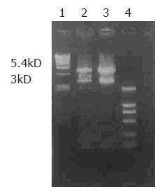
Figure 1 Restriction enzyme digestion of recombinant plasmids pcDNA3-Kit-W and pcDNA3-Kit-NW at XbaI and HindIII.
Lane 1: DNA/EcoRI+HindIII marker; lane 2: pcDNA3-Kit-W clone; lane 3: pcDNA3-Kit-NW clone; lane 4: DL-2000 marker.
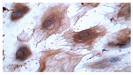
Figure 2 Positive immunocytochemical staining of C-kit transfected with pcDNA3-Kit-NW (×400).
Figure 3 Western blotting of pcDNA3-Kit-W, pcDNA3-Kit-NW, and pcDNA3 on HEKC line.
Lanes 1-3: pcDNA3-Kit-NW; lanes 4-6: pcDNA3-Kit-W; lanes 7 and 8: pcDNA3.
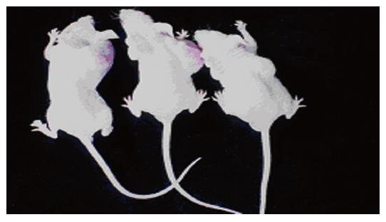
Figure 4 Tumor growth in nude mice 6 wk after being implanted with cells transfected with pcDNA3-Kit-NW.
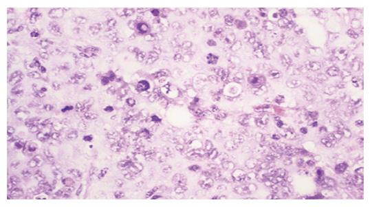
Figure 5 Giant, bizarre-shaped pyknotic nucleoli, or prominent pathologic mitosis in tumor (HE ×200).
- Citation: Bai CG, Liu XH, Xie Q, Feng F, Ma DL. A novel gain of function mutant in C-kit gene and its tumorigenesis in nude mice. World J Gastroenterol 2005; 11(45): 7104-7108
- URL: https://www.wjgnet.com/1007-9327/full/v11/i45/7104.htm
- DOI: https://dx.doi.org/10.3748/wjg.v11.i45.7104


