Copyright
©2005 Baishideng Publishing Group Inc.
World J Gastroenterol. Dec 7, 2005; 11(45): 7063-7071
Published online Dec 7, 2005. doi: 10.3748/wjg.v11.i45.7063
Published online Dec 7, 2005. doi: 10.3748/wjg.v11.i45.7063
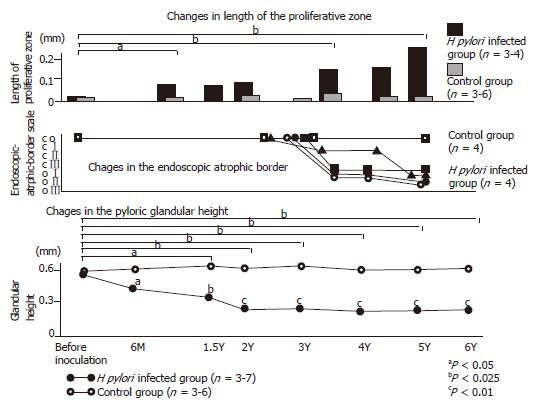
Figure 1 Gastric mucosal alteration of Japanese monkey model with H pylori infection.
Upper graph showed the gradual increase of the proliferative zone of H pylori-infected Japanese monkey model. Middle graph showed the alteration of endoscopic-atrophic-border scale of this model. Macroscopically, gastric atrophy advanced for more than 3 yr. Lower graph showed the alteration of the pyloric glandular height. Six months after inoculation, the pyloric glandular height was apparently lower in the infected animals than in controls. Furthermore, the atrophic change advanced gradually throughout the 6-yr observation period.
Figure 2 Microscopic views of the gastric body of Mongolian gerbils at 18 mo after H pylori inoculation.
A: Severe infiltration of polymorphonuclear and mononuclear cells were seen in the lamina propria. (HE stain, x100); B: Some glands have extended into the submucosa but not into the proper muscularis layer. Severe infiltration of mononuclear cells in the submucosa (HE stain, x10); C: Intestinal metaplasia is seen scattering in gastric mucosa (HE stain, x10); D: Intestinal metaplasia (Alcian blue stain (pH 2.5); original magnification, x10).
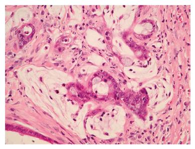
Figure 3 Microscopic views of the gastric mucosa of Mongolian gerbils at 18 mo after H pylori inoculation.
Well-differentiated adenocarcinoma has extended into the muscular layer. Atypical glands and nuclei and abnormal mitosis are evident (HE stain, x40).
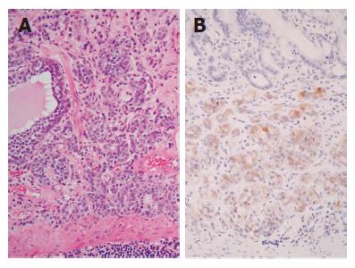
Figure 4 Gastric carcinoid in the stomach of a Mongolian gerbils colonized for 24 mo by H pylori.
Microscopic view showing intramucosal carcinoid tumor (A: HE stain, x20; B: immunohistochemistry of chromogranin A, x20’).
- Citation: Kodama M, Murakami K, Sato R, Okimoto T, Nishizono A, Fujioka T. Helicobacter pylori-infected animal models are extremely suitable for the investigation of gastric carcinogenesis. World J Gastroenterol 2005; 11(45): 7063-7071
- URL: https://www.wjgnet.com/1007-9327/full/v11/i45/7063.htm
- DOI: https://dx.doi.org/10.3748/wjg.v11.i45.7063


