Copyright
©2005 Baishideng Publishing Group Inc.
World J Gastroenterol. Nov 21, 2005; 11(43): 6888-6890
Published online Nov 21, 2005. doi: 10.3748/wjg.v11.i43.6888
Published online Nov 21, 2005. doi: 10.3748/wjg.v11.i43.6888
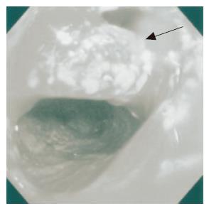
Figure 1 Pouchoscopy shows inflamed hemorrhagic mucosa.
An apical bridge is seen (arrow head).
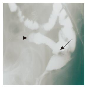
Figure 2 Pouchography shows a blind loop 10 cm in length (arrow head) and apical bridge formation (arrow).
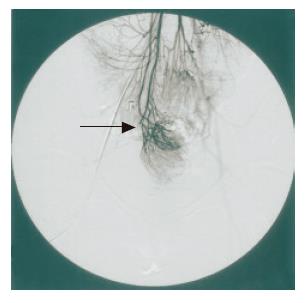
Figure 3 Superior mesenteric arteriography shows a marginal disconnection of the arcade (superior mesenteric artery–ileocolic artery) (arrow).
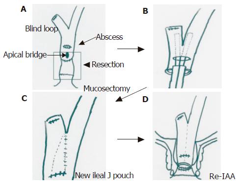
Figure 4 A: Mucosectomy and ileal pouch excision; B: side to side anastomosis of blind loop by linear stapler; C: reconstruction of a new ileal pouch; D: re-ileal pouch anal anastomosis.
- Citation: Toiyama Y, Araki T, Yoshiyama S, Miki C, Kusunoki M. Secondary pouchitis in a post-operative patient with ulcerative colitis, successfully treated by salvage surgery. World J Gastroenterol 2005; 11(43): 6888-6890
- URL: https://www.wjgnet.com/1007-9327/full/v11/i43/6888.htm
- DOI: https://dx.doi.org/10.3748/wjg.v11.i43.6888


