Copyright
©2005 Baishideng Publishing Group Inc.
World J Gastroenterol. Nov 21, 2005; 11(43): 6867-6870
Published online Nov 21, 2005. doi: 10.3748/wjg.v11.i43.6867
Published online Nov 21, 2005. doi: 10.3748/wjg.v11.i43.6867
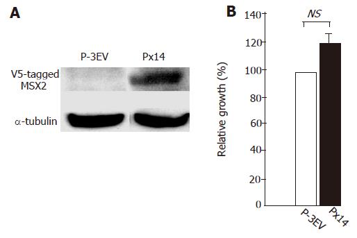
Figure 1 A: Western blotting analysis of V5-tagged MSX2 expression in Px14 cells; B: detection of cell proliferation of P-3EV and Px14 by BrdU incorporation assay.
No significant difference was observed in these two cell lines, but slight increase of proliferation ratio was observed in Px14 cells (unpaired t-test, n = 3).
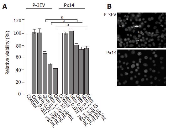
Figure 2 A: Cell viability of P-3EV and Px14 cell lines after 24 h of gemcitabine treatment at various concentrations.
Gemcitabine reduced cell viability in a dose-dependent manner. At a concentration of 0.1 μg/mL or above, Px14 cells exhibited higher viability relative to P-3EV cells (one-way ANOVA, n = 3, aP<0.05 vs others); B: Hoechst staining of P-3EV and Px14 cell lines. After 24 h of gemcitabine treatment at a concentration of 1 μg/mL, apoptotic changes (nuclear chromatin condensation and nuclear fragmentation) were obviously observed in P-3EV cells (arrow), but not in Px14 cells.
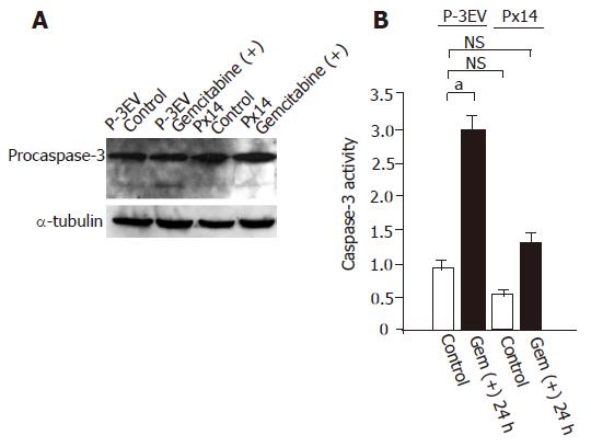
Figure 3 A: Western blotting analysis for caspase-3 activation.
Cells were harvested 24 h after gemcitabine (1 g/mL) or vehicle administration. In P-3EV cells, active caspase-3 (cleaved form) was detected after gemcitabine treatment (arrow), whereas in Px14 cells, cleaved form of caspase-3 was undetectable with or without gemcitabine; B: direct caspase-3 activity measurements by APOCYTO Caspase-3 Colorimetric Assay Kit. Gemcitabine (1 μg/mL) treatment significantly enhanced caspase-3 specific activity in P-3EV cells, but not in Px14 cells. No significant differences in caspase-3 activities were detected between vehicle-treated P-3EV cells and Px14 cells, even after gemcitabine treatment (one-way ANOVA, n = 3, aP<0.05 vs others).
- Citation: Hamada S, Satoh K, Kimura K, Kanno A, Masamune A, Shimosegawa T. MSX2 overexpression inhibits gemcitabine-induced caspase-3 activity in pancreatic cancer cells. World J Gastroenterol 2005; 11(43): 6867-6870
- URL: https://www.wjgnet.com/1007-9327/full/v11/i43/6867.htm
- DOI: https://dx.doi.org/10.3748/wjg.v11.i43.6867


