Copyright
©2005 Baishideng Publishing Group Inc.
World J Gastroenterol. Jan 28, 2005; 11(4): 557-560
Published online Jan 28, 2005. doi: 10.3748/wjg.v11.i4.557
Published online Jan 28, 2005. doi: 10.3748/wjg.v11.i4.557
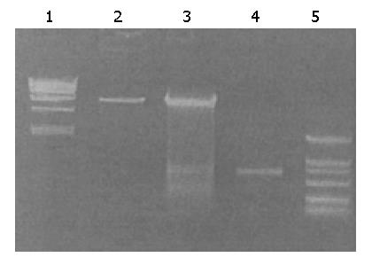
Figure 1 pcDNA 3HCV-Fc vectors identified by restriction enzyme analysis.
Lane 1: Marker (Ecoli); lane 2: pcDNA3; lane 3: pcDNA 3HCV-Fc was cut by restriction enzyme Xho I/Xba I; lane 4: Fc cDNA fragment was cloned into the vector; lane 5: Marker (DL-2000).
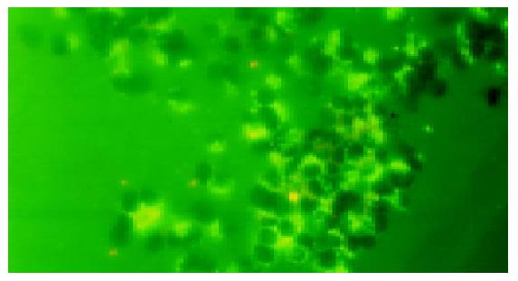
Figure 2 Expression of HCV C-Fc fusion proteins.
Figure 3 BM-derived DCs.
A: BM-derived DC (SEM×500); B: BM-derived DC (SEM×700).
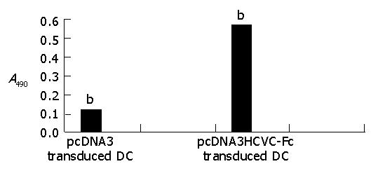
Figure 4 Serum anti-HCV C level in Balb/c mice.
bP<0.01 vs pcDNA3
- Citation: Wang QC, Feng ZH, Zhou YX, Nie QH. Induction of hepatitis C virus-specific cytotoxic T and B cell responses by dendritic cells expressing a modified antigen targeting receptor. World J Gastroenterol 2005; 11(4): 557-560
- URL: https://www.wjgnet.com/1007-9327/full/v11/i4/557.htm
- DOI: https://dx.doi.org/10.3748/wjg.v11.i4.557


