Copyright
©The Author(s) 2005.
World J Gastroenterol. Oct 21, 2005; 11(39): 6144-6151
Published online Oct 21, 2005. doi: 10.3748/wjg.v11.i39.6144
Published online Oct 21, 2005. doi: 10.3748/wjg.v11.i39.6144
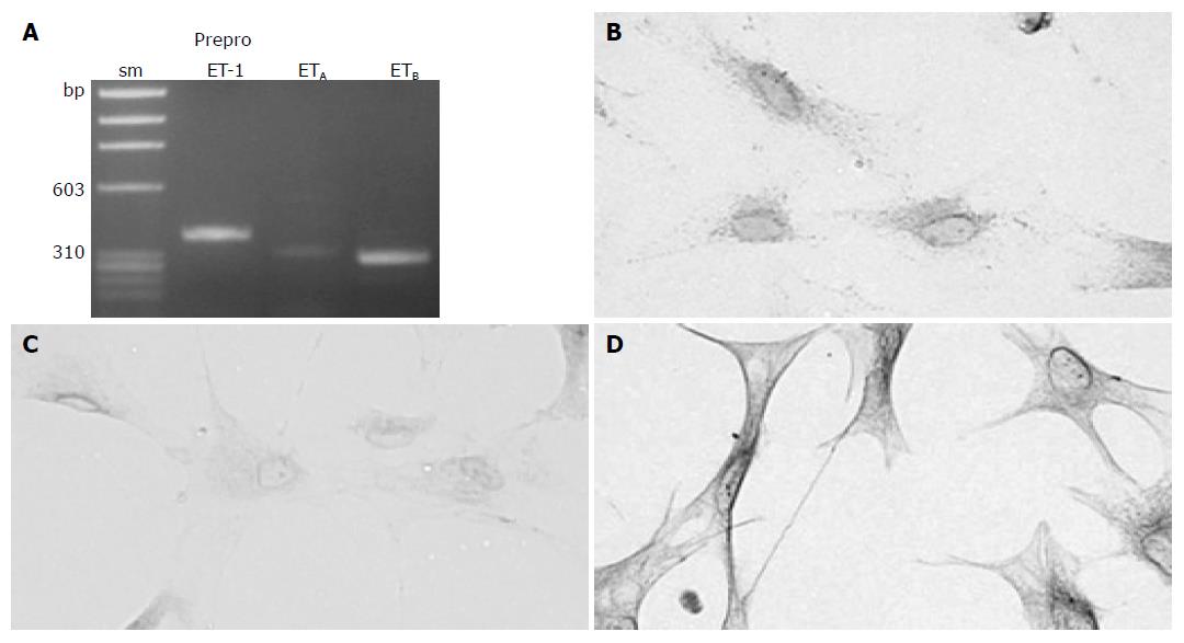
Figure 1 Culture-activated PSCs expressed ET and ET receptors.
A: Total RNA was prepared from culture-activated, serum-starved PSCs. The expression of preproET-1, ETA receptor and ETB receptor was examined by reverse transcription-PCR. The expected sizes of the PCR products were 382 bp for preproET-1, 321 bp for ETA receptor, and 297 bp for ETB receptor. sm: size marker (fX174 HaeIII digest). bp: bp; B-D: Serum-starved PSCs were grown directly on slides. Immunostaining for ET-1 (panel B), ETA receptor (panel C), and ETB receptor (panel D) was performed using a streptavidin-biotin peroxidase complex detection kit. Original magnification: ×0 objective.
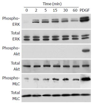
Figure 2 ET-1 induced phosphorylation of ERK and MLC, but not Akt.
PSCs were treated with ET-1 (at 100 nmol/L) for the indicated time. Total cell lysates (approximately 100 mg) were prepared, and the levels of phosphorylated ERK, Akt, and MLC were determined by Western blotting. As positive controls, total cell lysates were prepared from PSCs treated with PDGF-BB (at 25 mg/L) for 5 min, and Western blotting was performed in a similar manner. Levels of total ERK, Akt, and MLC were also determined.
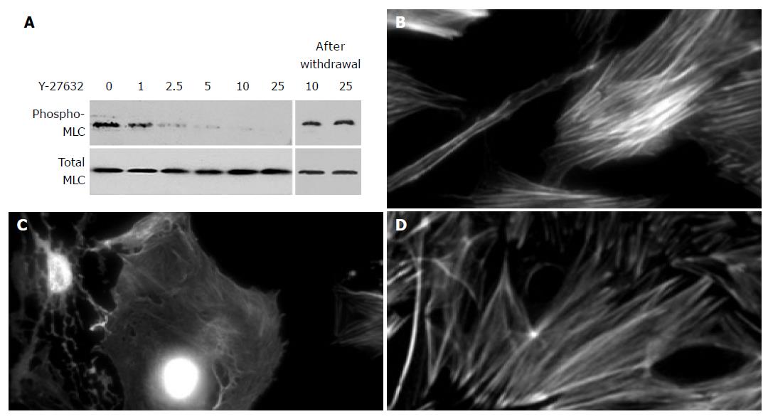
Figure 3 Y-27632 decreased stress fiber formation.
A: PSCs were treated with ET-1 (at 100 nmol/L) in the presence of Y-27632 at the indicated concentrations (at mmol/L) for 30 min. Total cell lysates (approximately 100 mg) were prepared, and the levels of phosphorylated and total MLC were determined by Western blotting. In parallel experiments, Y-27632 was withdrawn after 30-min incubation, and replaced with 10% FBS. After 48-h incubation with 10% FBS, total cell lysates were prepared, and the levels of phosphorylated and total MLC were determined by Western blotting; B-D: PSCs were incubated with ET-1 (at 100 nmol/L) in the absence (B) or presence (C) of Y-27632 (at 25 mmol/L) for 24 h, and stained with rhodamine-labeled phalloidin. In parallel experiments, Y-27632 was withdrawn and replaced with 10% FBS. After 48-h incubation with 10% FBS, PSCs were stained with rhodamine-labeled phalloidin (D). Original magnification: ×0 objective.
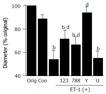
Figure 4 ET-1 induced contraction of PSCs.
PSCs were plated on the surface of collagen lattices prepared in each well of a six-well plate and incubated for 2 h to allow adherence. The collagen lattices were then detached from the plate by gentle circumferential dislodgment. Serum-starved PSCs were left untreated ("Con" or were treated with ET-1 (at 100 nmol/L) in the absence or presence of BQ-123 (×23×at 10 mmol/L), BQ-788 (×88×at 10 mmol/L), Y-27632 (at 25 mmol/L), or U0126 ("U"at 5 mmol/L). Lattices were then detached from the plate, and incubated in serum-free medium (control, "Con" with or without 100 nmol/L ET-1. After 24-h incubation, we measured the diameter of the collagen lattices, and calculated the × of the original diameter ("Orig"= 35 mm)× Data are shown as mean±SD (% of the original diameter, n = 8). bP<0.01 vs control (serum-free medium only), dP<0.01 vs ET-1 treatment only.
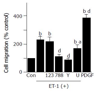
Figure 5 ET-1 induced migration of PSCs.
Cell migration was assessed using modified Boyden chambers with 8-mm pore filters. Serum-starved PSCs were left untreated ("Con" or were treated with ET-1 (at 100 nmol/L) in the lower chamber in the absence or presence of BQ-123 (×23×at 10 mmol/L), BQ-788 (×88×at 10 mmol/L), Y-27632 (at 25 mmol/L), or U0126 ("U"at 5 mmol/L). After 24-h incubation, the cells migrated to the underside of the filter were stained, and counted under a light microscopy. As positive controls, cells were treated with PDGF-BB (at 25 mg/L) in the lower chamber, and cell migration was assessed in a similar manner. Data are shown as mean±SD (% of the control, n = 6). bP<0.01 vs control (serum-free medium only), aP<0.05, dP<0.01 vs ET-1 treatment only.
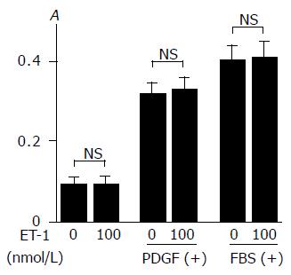
Figure 6 ET-1 did not alter basal or inducible proliferation of PSCs.
Serum-starved PSCs were treated with ET-1 at 0 or 100 nmol/L in the absence or presence of PDGF-BB (at 25 mg/L) or 100 mL/L fetal bovine serum ("FBS". After 24-h incubation, DNA synthesis was assessed by BrdU incorporation enzyme-linked immunosorbent assay. Data are shown as mean±SD (n = 6). NS: not significant, A: optical density.
- Citation: Masamune A, Satoh M, Kikuta K, Suzuki N, Satoh K, Shimosegawa T. Endothelin-1 stimulates contraction and migration of rat pancreatic stellate cells. World J Gastroenterol 2005; 11(39): 6144-6151
- URL: https://www.wjgnet.com/1007-9327/full/v11/i39/6144.htm
- DOI: https://dx.doi.org/10.3748/wjg.v11.i39.6144


