Copyright
©The Author(s) 2005.
World J Gastroenterol. Oct 7, 2005; 11(37): 5807-5810
Published online Oct 7, 2005. doi: 10.3748/wjg.v11.i37.5807
Published online Oct 7, 2005. doi: 10.3748/wjg.v11.i37.5807
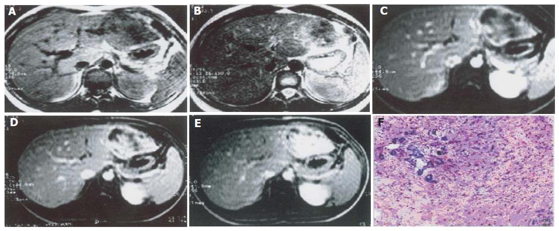
Figure 1 Solid chief dingle mass type of MHL on T1WI(A), T2WI(B), and dynamic enhancement in artery phase(C), portal rein phase(D), and latency phase(E), and its pathologic lesions(F).
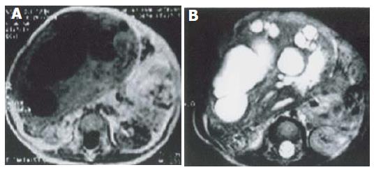
Figure 2 Mixed cystic-solid single mass type of MHL on T1WI(A) and T2WI(B).
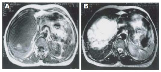
Figure 3 Cystic single mass type of MHL on T1WI(A), T2WI(B).
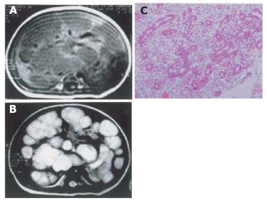
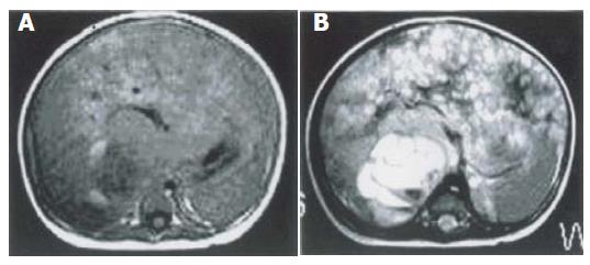
Figure 4 Diffuse and multifocal type of MHL on T1WI(A), T2WI(B) and Pathologic picture(C).
Figure 5 Diffuse and single combined type of MHL on T1WI(A) and T2WI(B).
- Citation: Ye BB, Hu B, Wang LJ, Liu HS, Zou Y, Zhou YB, Kang Z. Mesenchymal hamartoma of liver: Magnetic resonance imaging and histopathologic correlation. World J Gastroenterol 2005; 11(37): 5807-5810
- URL: https://www.wjgnet.com/1007-9327/full/v11/i37/5807.htm
- DOI: https://dx.doi.org/10.3748/wjg.v11.i37.5807


