Copyright
©The Author(s) 2005.
World J Gastroenterol. Oct 7, 2005; 11(37): 5801-5806
Published online Oct 7, 2005. doi: 10.3748/wjg.v11.i37.5801
Published online Oct 7, 2005. doi: 10.3748/wjg.v11.i37.5801
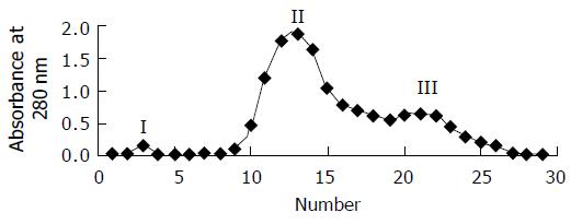
Figure 1 Chromatography of soybean conglycinin on sepharose-CL-6B.
Bed size,1 cmx100 cm, flow rate 30 mL/h, absorbance at 280 nm.
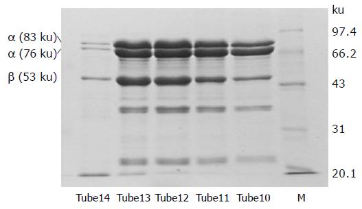
Figure 2 SDS-PAGE pattern of purified conglycinin.
a, a and b indicate subunits of conglycinin. M: standard molecular weight proteins.
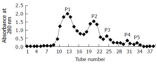
Figure 3 Chromatography of soybean conglycinin hydrolysates on sephadex-G15.
Bed size, 1.0 cmx100 cm, flow rate 15 mL/h, absorbance at 280 nm.
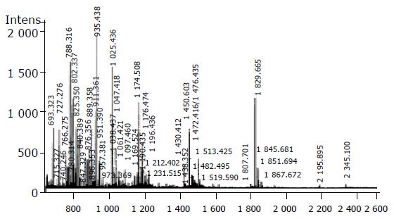
Figure 4 MALDI-TOF-MS analysis of fraction P2.
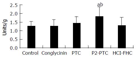
Figure 5 Effect of conglycinin peptides on b-galactosidase activities in cecal contents (n = 8).
Values are expressed as the mean±SD, aP < 0.05 vs control group, bP < 0.01 vs conglycinin group.
-
Citation: Zuo WY, Chen WH, Zou SX. Separation of growth-stimulating peptides for
Bifidobacterium from soybean conglycinin. World J Gastroenterol 2005; 11(37): 5801-5806 - URL: https://www.wjgnet.com/1007-9327/full/v11/i37/5801.htm
- DOI: https://dx.doi.org/10.3748/wjg.v11.i37.5801


