Copyright
©The Author(s) 2005.
World J Gastroenterol. Oct 7, 2005; 11(37): 5770-5776
Published online Oct 7, 2005. doi: 10.3748/wjg.v11.i37.5770
Published online Oct 7, 2005. doi: 10.3748/wjg.v11.i37.5770
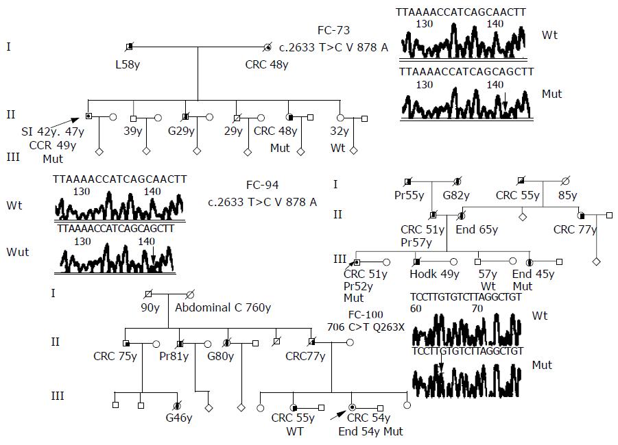
Figure 1 Pedigrees and DNA nucleotide sequence of the three families with disease-causing mutations of hMSH6 gene.
Squares, males; circles, females; diagonal bars, deceased; unblackened symbols, no tumor; semi-blackened symbols, patients with histological-verified carcinomas; arrows, probands; mut, carries the indicated mutation. Abbreviations indicating type of tumor: CRC, colorectal; SI, small intestine; G, gastric; H, Hodgkin; End, endometrium; Pr, prostate; L, liver. Number after abbreviation indicates age at which tumor was diagnosed. Electropherograms of the wild type and mutant nucleotide sequences for V878A and Q263X mutations located in exon 4 of the hMSH6 gene.
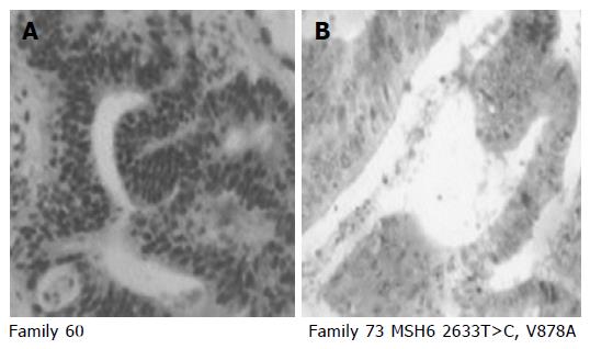
Figure 2 Representative examples of positive (A) and negative (B) immunohistochemical staining for hMSH6 protein (clone 44, Transduction laboratories/Becton Dickinson, Lexington, UK).
A: Tumor from family 60, exhibited positive nuclear staining for hMSH6; B: tumor from family 73 carrier of V878A mutation, exhibited loss of hMSH6 expression.
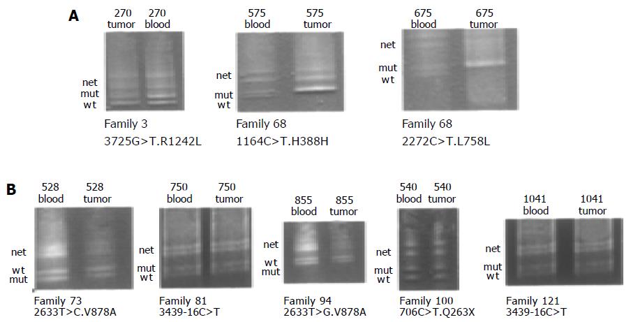
Figure 3 DGGE-LOH analysis of different hMSH6 exons, amplified from both tumor and blood DNA.
Analysis of 7 patients (270, 675, 526, 750, 855, 940, and 1 041) harboring the mutations R1242L, H388L, L758L, V878A, 3439-16 C > T, and Q263X. Bands corresponding to wild-type (Wt), mutant (Mut) alleles and the heteroduplex (Het) are indicated in all cases. Panel A shows LOH of the mutant allele in patient 270 and LOH of wild-type allele in patient 675. Panel B shows retention of both alleles (wt and mut) in all patients.
- Citation: Abajo AS, Hoya ML, Tosar A, Godino J, Fernández JM, Asenjo JL, Villamil BP, Segura PP, Diaz-Rubio E, Caldes T. Low prevalence of germline hMSH6 mutations in colorectal cancer families from Spain. World J Gastroenterol 2005; 11(37): 5770-5776
- URL: https://www.wjgnet.com/1007-9327/full/v11/i37/5770.htm
- DOI: https://dx.doi.org/10.3748/wjg.v11.i37.5770


