Copyright
©The Author(s) 2005.
World J Gastroenterol. Sep 28, 2005; 11(36): 5742-5745
Published online Sep 28, 2005. doi: 10.3748/wjg.v11.i36.5742
Published online Sep 28, 2005. doi: 10.3748/wjg.v11.i36.5742
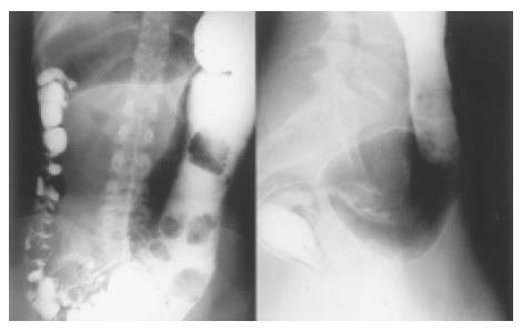
Figure 1 Contrast medium enema of the distal bowel shows a distal stenosis and a megacolon proximal to the stenosis.
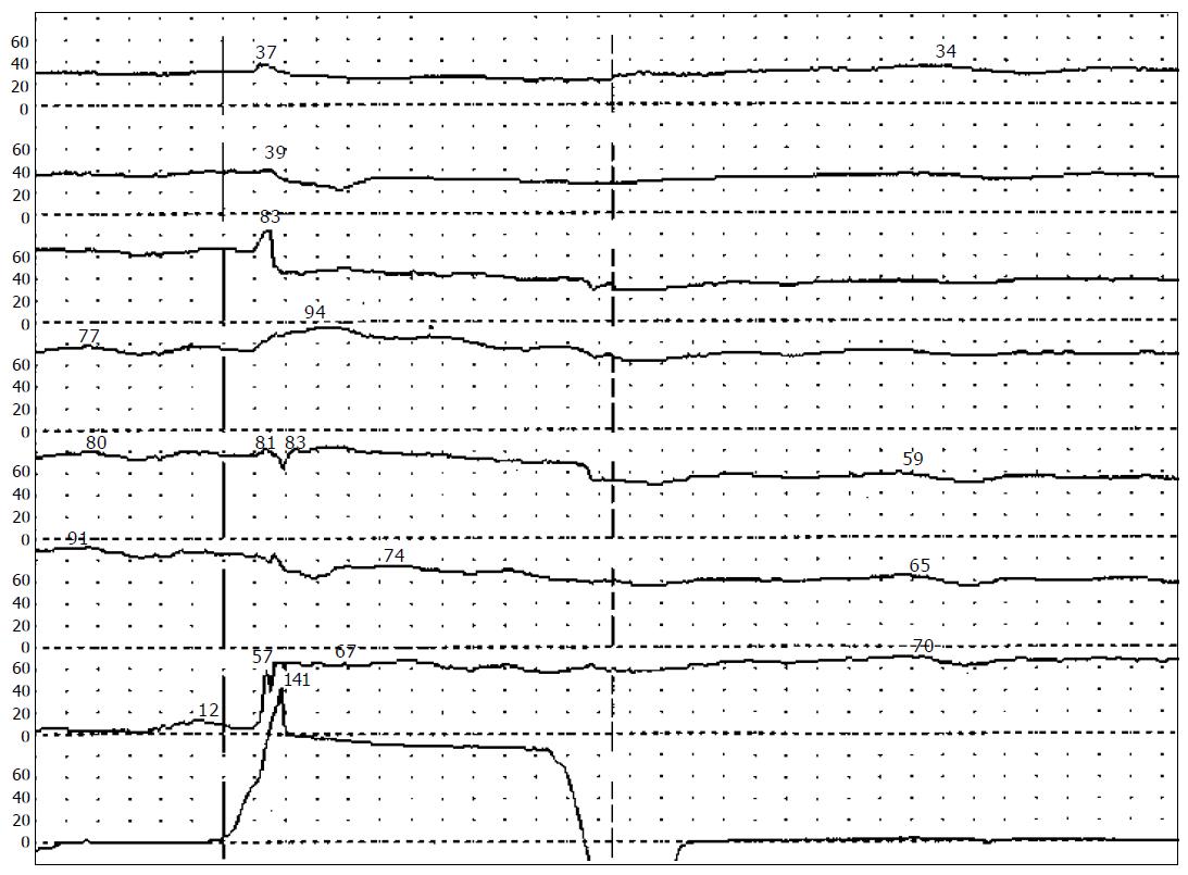
Figure 2 Anorectal manometry shows absence of the recto-anal inhibitory reflex after inflation of an intrarectally placed balloon with 40 mL of air.
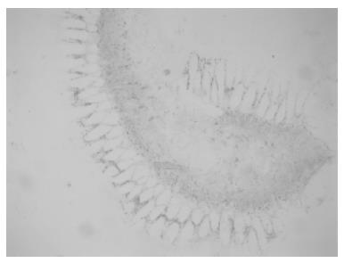
Figure 3 Rectal biopsy (AChE-staining) shows increased activity of acetylcholinesterase in the mucosal layer of the dilated segment.
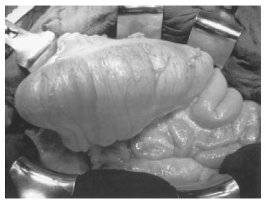
Figure 4 Megacolon in situ.
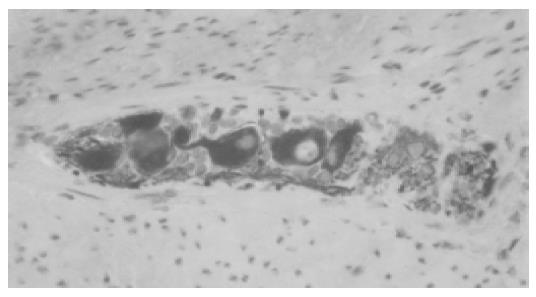
Figure 5 Instantaneous section (Peripherin-immmunohistochemistry) shows normal formation of plexus myentericus.
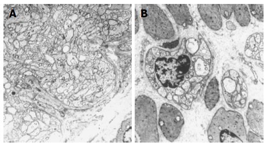
Figure 6 Electron microscopic sections show thickened parasympathetic nerve fibers in the distal part of the resected segment (A) and normal numbers of ganglia in the proximal part (B).
- Citation: Werner CR, Stoltenburg-Didinger G, Weidemann H, Benckert C, Schmidtmann M, Voort IRVD, Andresen V, Klapp BF, Neuhaus P, Wiedenmann B, Mönnikes H. Megacolon in adulthood after surgical treatment of Hirschsprung’s disease in early childhood. World J Gastroenterol 2005; 11(36): 5742-5745
- URL: https://www.wjgnet.com/1007-9327/full/v11/i36/5742.htm
- DOI: https://dx.doi.org/10.3748/wjg.v11.i36.5742


