Copyright
©The Author(s) 2005.
World J Gastroenterol. Sep 28, 2005; 11(36): 5659-5665
Published online Sep 28, 2005. doi: 10.3748/wjg.v11.i36.5659
Published online Sep 28, 2005. doi: 10.3748/wjg.v11.i36.5659
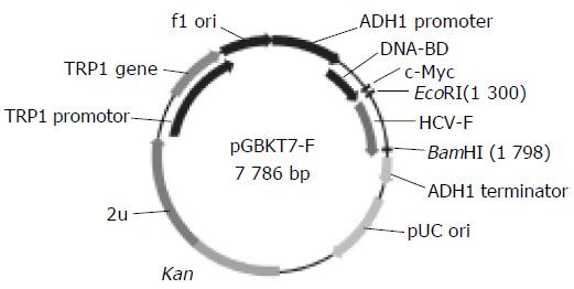
Figure 1 Map of “bait” plasmid pGBKT7-F.
Figure 2 Sequences of PCR products A: a 486-bp fragment-F amplified by amplified by RT-PCR; D1: pGBKT7-FBP2; D2: pGADT7-FBP2 cut by EcoRI/RT-PCR; B: pGBKT7-F cut by EcoRI/BamHI; C: a 840-bp fragment-FBP2 BamHI.
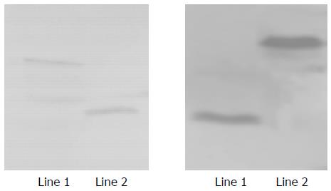
Figure 3 Expression of HCV F and HCV FBP2 proteins in yeast.
Lane 1: HCV F protein; lane 2: positive control; lane 3: HCV FBP2 protein.
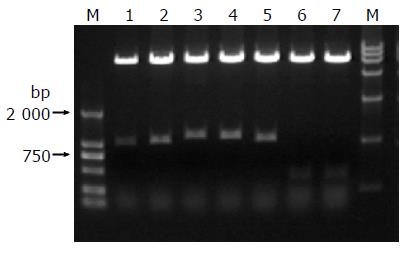
Figure 4 Identification of different colonies by BglII digestion.
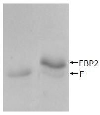
Figure 5 Interaction between HCV F protein and FBP2 protein identified by co-immunoprecipitation.
Lane 1: HCV F protein; lane 2: interaction between HCV F and FBP2 proteins.
- Citation: Huang YP, Cheng J, Zhang SL, Wang L, Guo J, Liu Y, Yang Y, Zhang LY, Bai GQ, Gao XS, Ji D, Lin SM, Shao Q. Screening of hepatocyte proteins binding to F protein of hepatitis C virus by yeast two-hybrid system. World J Gastroenterol 2005; 11(36): 5659-5665
- URL: https://www.wjgnet.com/1007-9327/full/v11/i36/5659.htm
- DOI: https://dx.doi.org/10.3748/wjg.v11.i36.5659


