Copyright
©2005 Baishideng Publishing Group Inc.
World J Gastroenterol. Sep 21, 2005; 11(35): 5475-5479
Published online Sep 21, 2005. doi: 10.3748/wjg.v11.i35.5475
Published online Sep 21, 2005. doi: 10.3748/wjg.v11.i35.5475
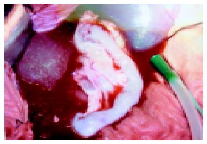
Figure 1 Pale pancreas after perfusion.
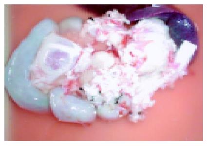
Figure 2 Modified graft for transplantation.
Figure 3 Anastomosis of donor portal vein to recipient superior mesenteric vein (A), donor common iliac artery to recipient aorta (B) in an end-to-side fashion, and donor duodenum to recipient jejunum (C) in a side-to-side fashion.
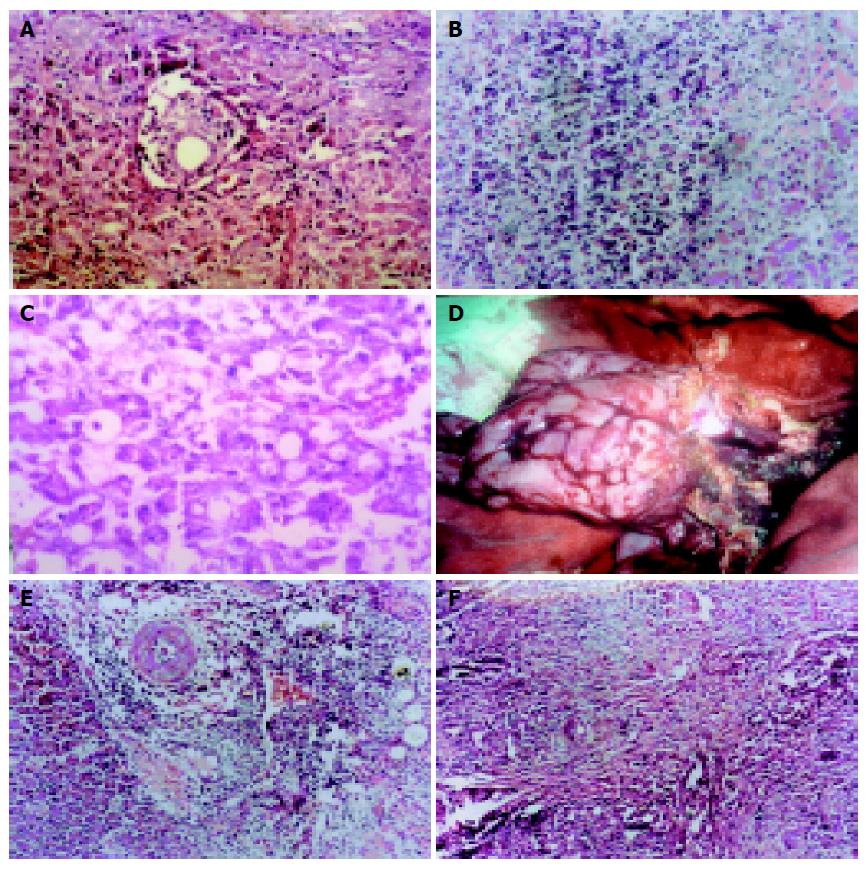
Figure 4 Mild rejection 3 d after operation (A) (HE ×200) and moderate rejection 5 d after operation (B) (HE ×100), apoptosis body and pancreatic edema 7 d after operation (C), pale pancreas with flexible mild edema 9 d after operation (D), severe rejection 10 d after operation (E), and infiltration of lymphocytes and hyperplasia of connective tissue 21 d after operation (F).
- Citation: Zhang ZD, Han FH, Meng LX. Establishment of a pig model with enteric and portal venous drainage of pancreatoduodenal transplantation. World J Gastroenterol 2005; 11(35): 5475-5479
- URL: https://www.wjgnet.com/1007-9327/full/v11/i35/5475.htm
- DOI: https://dx.doi.org/10.3748/wjg.v11.i35.5475


