Copyright
©The Author(s) 2005.
World J Gastroenterol. Sep 14, 2005; 11(34): 5416-5417
Published online Sep 14, 2005. doi: 10.3748/wjg.v11.i34.5416
Published online Sep 14, 2005. doi: 10.3748/wjg.v11.i34.5416
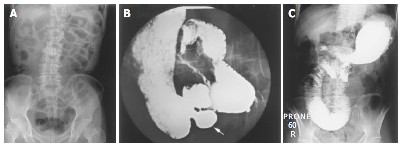
Figure 1 A plain X-ray of the abdomen showing dilatation of the small intestine, especially the proximal jejunum (A).
B and C: An upper GI oral contrast study showing multiple diverticula in the duodenum and proximal jejunum (the white arrow, (B) and dilatation of the proximal jejunum (C).
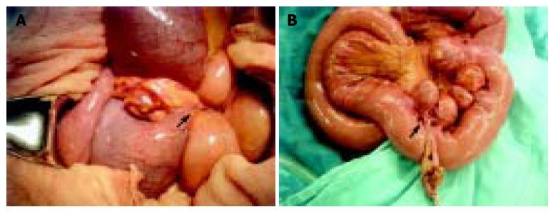
Figure 2 The photograph shows an adhesion band overriding the jejunum (the black arrow), with ischemic changes in the proximal jejunum (A).
After surgical removal of the adhesion band, we found that the band was connected to one of the diverticula, arising from the mesentery side (the black arrow) (B).
- Citation: Lin CH, Hsieh HF, Yu CY, Yu JC, Chan DC, Chen TW, Chen PJ, Liu YC. Diverticulosis of the jejunum with intestinal obstruction: A case report. World J Gastroenterol 2005; 11(34): 5416-5417
- URL: https://www.wjgnet.com/1007-9327/full/v11/i34/5416.htm
- DOI: https://dx.doi.org/10.3748/wjg.v11.i34.5416


