Copyright
©The Author(s) 2005.
World J Gastroenterol. Sep 14, 2005; 11(34): 5394-5397
Published online Sep 14, 2005. doi: 10.3748/wjg.v11.i34.5394
Published online Sep 14, 2005. doi: 10.3748/wjg.v11.i34.5394
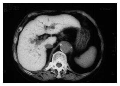
Figure 1 An unenhanced computed tomography (CT) scan of the upper abdomen.
The liver has a greatly increased density (111.8 HU).
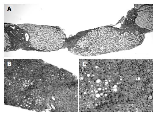
Figure 2 Microscopic features of the liver biopsy specimen.
A: Micronodular cirrhosis has become established (silver impregnation, scale bar = 200 µm); B: Low power view of the liver biopsy specimen (hematoxylin and eosin stain, scale bar = 200 µm). Fatty degeneration and marked fibrosis with inflammatory cell infiltration were observed; C: High power view of the liver biopsy specimen (hematoxylin and eosin stain, scale bar = 20 µm). Foci of hepatocellular necrosis and dense fibrosis with neutrophilic infiltration were observed. Hepatocytes showed both the microvesicular and macrovesicular types of steatosis.
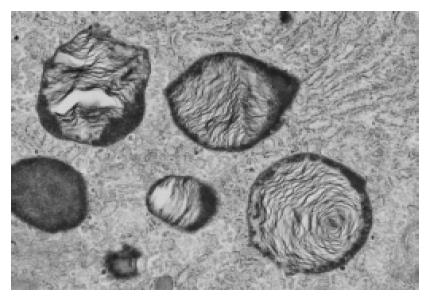
Figure 3 Transmission electron micrograph of the biopsy specimen with lamellated inclusions in the cytoplasm of the hepatocyte (original magnification ×20 000).
- Citation: Oikawa H, Maesawa C, Sato R, Oikawa K, Yamada H, Oriso S, Ono S, Yashima-Abo A, Kotani K, Suzuki K, Masuda T. Liver cirrhosis induced by long-term administration of a daily low dose of amiodarone: A case report. World J Gastroenterol 2005; 11(34): 5394-5397
- URL: https://www.wjgnet.com/1007-9327/full/v11/i34/5394.htm
- DOI: https://dx.doi.org/10.3748/wjg.v11.i34.5394


