Copyright
©The Author(s) 2005.
World J Gastroenterol. Sep 14, 2005; 11(34): 5314-5321
Published online Sep 14, 2005. doi: 10.3748/wjg.v11.i34.5314
Published online Sep 14, 2005. doi: 10.3748/wjg.v11.i34.5314
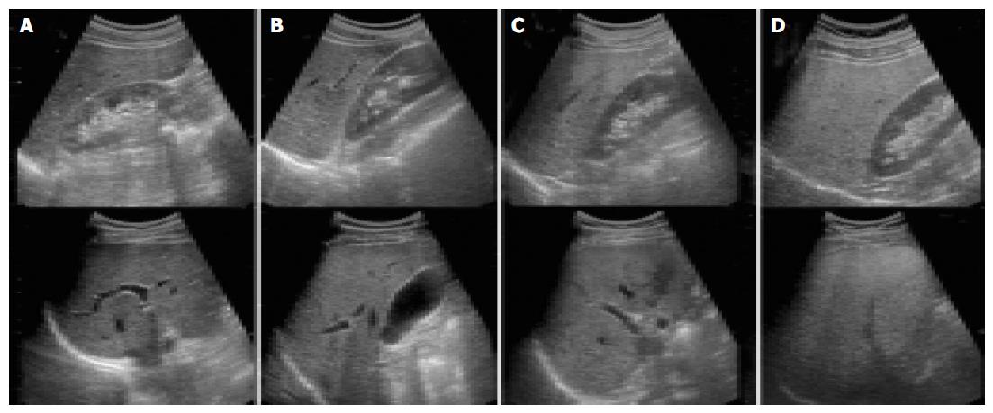
Figure 1 Ultrasonographic findings of liver according to the degree of steatosis.
(A) Normal liver, (B) mild steatosis: a slight augmentation in liver echogenicity, a slight exaggeration of echogenic discrepancy between liver and kidney, and a relative preservation of echo line in the portal vein wall. (C) Moderate steatosis: a loss of echo line in the portal vein wall, particularly from the peripheral branches, resulting in a featureless appearance of the liver. (D) Severe steatosis: much greater reduction in echo penetration, a loss of echogenicity in most of the portal vein wall, including the main branches, and a large echo discrepancy between liver and kidney. Adapted from Saverymuttu et al[17].
- Citation: Kim HC, Choi SH, Shin HW, Cheong JY, Lee KW, Lee HC, Huh KB, Kim DJ. Severity of ultrasonographic liver steatosis and metabolic syndrome in Korean men and women. World J Gastroenterol 2005; 11(34): 5314-5321
- URL: https://www.wjgnet.com/1007-9327/full/v11/i34/5314.htm
- DOI: https://dx.doi.org/10.3748/wjg.v11.i34.5314


