Copyright
©The Author(s) 2005.
World J Gastroenterol. Sep 14, 2005; 11(34): 5277-5282
Published online Sep 14, 2005. doi: 10.3748/wjg.v11.i34.5277
Published online Sep 14, 2005. doi: 10.3748/wjg.v11.i34.5277
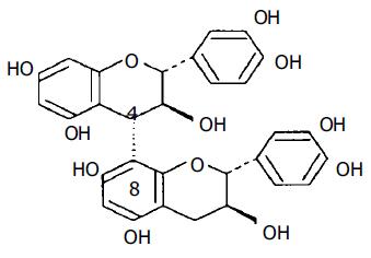
Figure 1 Chemical structure of procyanidin B3.
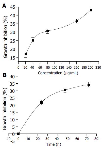
Figure 2 Dose-dependent effects (A) and time-dependent effects (B) of PMBE on inhibition of BEL-7402 cell growth.
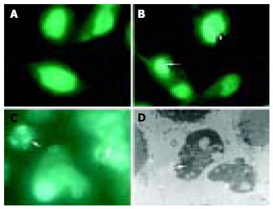
Figure 3 Morphological changes in BEL-7402 cells incubated without PMBE for 24 h (A), with 160 µg/mL PMBE for 24 (B), 48 (C) and 72 h (D).
Arrows denote condensed, fragmented nuclei, apoptotic bodies.
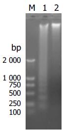
Figure 4 Apoptosis induced by PMBE.
M: DNA marker DL2000; 1: Treatment with PMBE; 2: Untreated negative control.
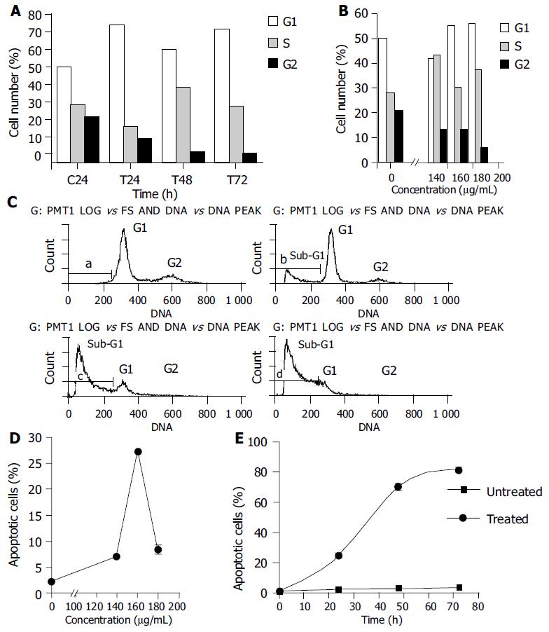
Figure 5 Time-course effects and dose–response effects of PMBE on cell cycle distribution (A and B) and apoptosis of BEL-7402 cells (C-E).
a: control 24 h; b: 24 h; c: 48 h; d: 72 h.
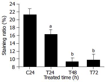
Figure 6 Expression of Bcl-2 protein in BEL-7402 cells treated with 160 µg/mL PMBE for different time periods.
C: control; T: treatment with PMBE; aP < 0.05 as significant, bP < 0.01 very significant vs the control.
- Citation: Cui YY, Xie H, Qi KB, He YM, Wang JF. Effects of Pinus massoniana bark extract on cell proliferation and apoptosis of human hepatoma BEL-7402 cells. World J Gastroenterol 2005; 11(34): 5277-5282
- URL: https://www.wjgnet.com/1007-9327/full/v11/i34/5277.htm
- DOI: https://dx.doi.org/10.3748/wjg.v11.i34.5277


