Copyright
©The Author(s) 2005.
World J Gastroenterol. Sep 7, 2005; 11(33): 5251-5253
Published online Sep 7, 2005. doi: 10.3748/wjg.v11.i33.5251
Published online Sep 7, 2005. doi: 10.3748/wjg.v11.i33.5251
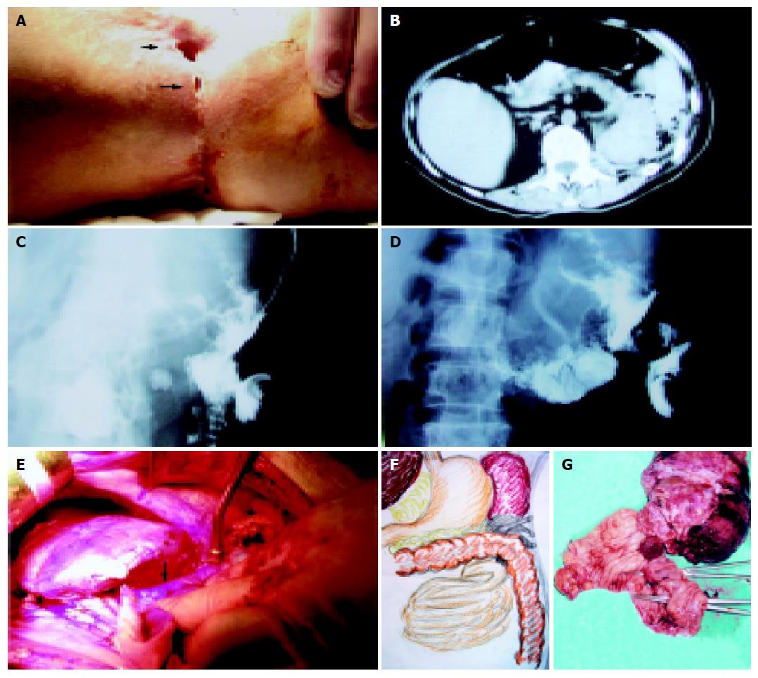
Figure 1 A: Surgical scar over left flank with two fistulous openings; B: Abdominal CT scan revealed irregular mass between the pancreatic tail, spleen and left lateral abdominal wall with air bubbles; C: The fistulogram revealed communication between the skin and sinus tract; D: The fistulogram revealed communication between the skin and intestinal tract; E: At laparotomy, splenomegaly and dense adhesion to the splenic flexure of colon (arrow) and the left abdominal wall were noted; F: The drawing revealed the relation between the fragile soft mass, the fistulas, the spleen and the bowels; G: The specimen was shown and these two openings in the colon communicating with skin were indexed with the instruments.
Figure 2 Microscopic photograph.
A: The squamous cancer cells and keratin pearls (arrow) were shown on the microscopic photograph; B: The pathological photograph revealed fistulous tract (arrow) and the tumor cells arose from its epithelium; C: The microscopic photograph revealed the tumor cells (arrow) invading the spleen (triangular arrow); D: The microscopic photograph revealed the tumor cells (arrow) invading the colon (triangular arrow).
- Citation: Lee YT, Hsu SD, Kuo CL, Chou DA, Lin MS, Huang MH, Wu HS. Squamous cell carcinoma arising from longstanding colocutaneous fistula: A case report. World J Gastroenterol 2005; 11(33): 5251-5253
- URL: https://www.wjgnet.com/1007-9327/full/v11/i33/5251.htm
- DOI: https://dx.doi.org/10.3748/wjg.v11.i33.5251


