Copyright
©The Author(s) 2005.
World J Gastroenterol. Sep 7, 2005; 11(33): 5248-5250
Published online Sep 7, 2005. doi: 10.3748/wjg.v11.i33.5248
Published online Sep 7, 2005. doi: 10.3748/wjg.v11.i33.5248
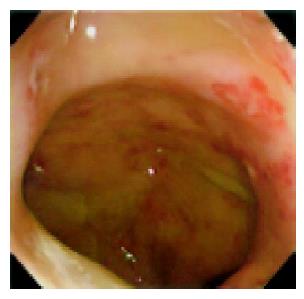
Figure 1 Colonoscopy showing multiple aphthous ulcers and erosion from the rectum to the ileocecal region.
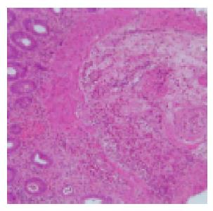
Figure 2 A colonic biopsy specimen showing marked cellular infiltration from the muscular layer of the mucosa to the submucosa and small vessels, lumen stenosis with thickening of the intima, indicative of vasculitis.
A H&E,×100.
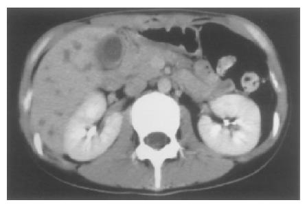
Figure 3 CT showing the gallbladder distended with a thickened wall.
In addition, low density areas faintly enhanced at S6 and S8 in the right lobe of the liver suspected to be abscesses are seen.
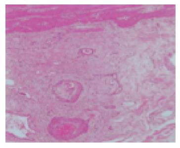
Figure 4 Histopathological findings of the resected gallbladder revealed necrotized vessels with occlusive intimal proliferation below the tunica serosa vesicae felleae, which had passed acute phase in the resected specimen.
We could not detect eosinophilic infiltration in this specimen.
- Citation: Suzuki M, Nabeshima K, Miyazaki M, Yoshimura H, Tagawa S, Shiraki K. Churg-Strauss syndrome complicated by colon erosion, acalculous cholecystitis and liver abscesses. World J Gastroenterol 2005; 11(33): 5248-5250
- URL: https://www.wjgnet.com/1007-9327/full/v11/i33/5248.htm
- DOI: https://dx.doi.org/10.3748/wjg.v11.i33.5248


