Copyright
©The Author(s) 2005.
World J Gastroenterol. Sep 7, 2005; 11(33): 5241-5244
Published online Sep 7, 2005. doi: 10.3748/wjg.v11.i33.5241
Published online Sep 7, 2005. doi: 10.3748/wjg.v11.i33.5241
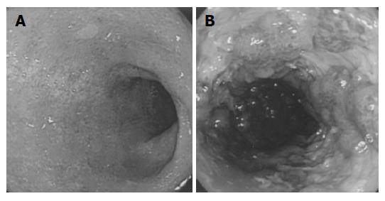
Figure 1 Diffuse mucosal edema, loss of vascular pattern, and friability without ulcerations in the rectum and distal sigmoid colon (A) and multiple longitudinal ulcers and pseudopolyps in the rectosigmoid colon (B).
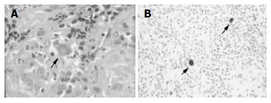
Figure 2 Enlarged histiocyte with Cowdry A type intranuclear inclusion body (A) and histiocytes positive for immunostaining of the CMV antigen (B).
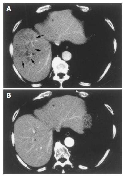
Figure 3 Small hypo-dense lesions in the right lobe of the liver, suggestive of multiple liver abscesses (A) and no evidence of residual hypo-dense lesions after treatment with meropenem (B).
- Citation: Inoue T, Hirata I, Egashira Y, Ishida K, Kawakami K, Morita E, Murano N, Yasumoto S, Murano M, Toshina K, Nishikawa T, Hamamoto N, Nakagawa K, Katsu KI. Refractory ulcerative colitis accompanied with cytomegalovirus colitis and multiple liver abscesses: A case report. World J Gastroenterol 2005; 11(33): 5241-5244
- URL: https://www.wjgnet.com/1007-9327/full/v11/i33/5241.htm
- DOI: https://dx.doi.org/10.3748/wjg.v11.i33.5241


