Copyright
©The Author(s) 2005.
World J Gastroenterol. Sep 7, 2005; 11(33): 5232-5234
Published online Sep 7, 2005. doi: 10.3748/wjg.v11.i33.5232
Published online Sep 7, 2005. doi: 10.3748/wjg.v11.i33.5232
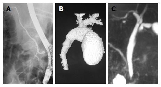
Figure 1 Reversed cholangiopancreatograms.
A: ERCP; B: 3D-DIC CT; C: MRCP.
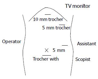
Figure 2 Arrangement around the operating table and positions of the trocars.
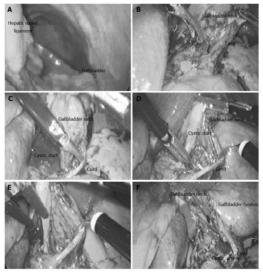
Figure 3 Laparoscopic view during cholecystectomy.
A: Gallbladder was placed on the left side and hepatic round ligament on the right; B: the fundus of gallbladder was grasped and tracted; C: the cord was revealed lying inferior to the cystic duct; D: the surroundings of the cystic duct were dissected after the cord was tracted; E: the cystic duct was cut after the clip ligature; F: the cord lead only to the gallbladder.
- Citation: Kamitani S, Tsutamoto Y, Hanasawa K, Tani T. Laparoscopic cholecystectomy in situs inversus totalis with “inferior” cystic artery: A case report. World J Gastroenterol 2005; 11(33): 5232-5234
- URL: https://www.wjgnet.com/1007-9327/full/v11/i33/5232.htm
- DOI: https://dx.doi.org/10.3748/wjg.v11.i33.5232


