Copyright
©The Author(s) 2005.
World J Gastroenterol. Sep 7, 2005; 11(33): 5226-5228
Published online Sep 7, 2005. doi: 10.3748/wjg.v11.i33.5226
Published online Sep 7, 2005. doi: 10.3748/wjg.v11.i33.5226
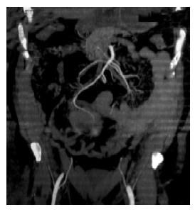
Figure 1 Abdominal CT scan showing a round, lobulated mass in the mesenteric region, tightly related to the bowel wall.
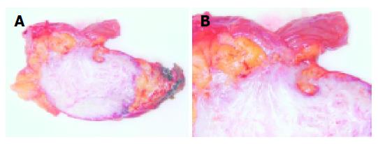
Figure 2 The resected small bowel with a well circumscribed firm mass in the mesenteric adipose tissue which exhibits an expanding growth pattern (A).
The tumor has a tan-gray appearance on cut surface, and focally infiltrates the bowel wall (B).
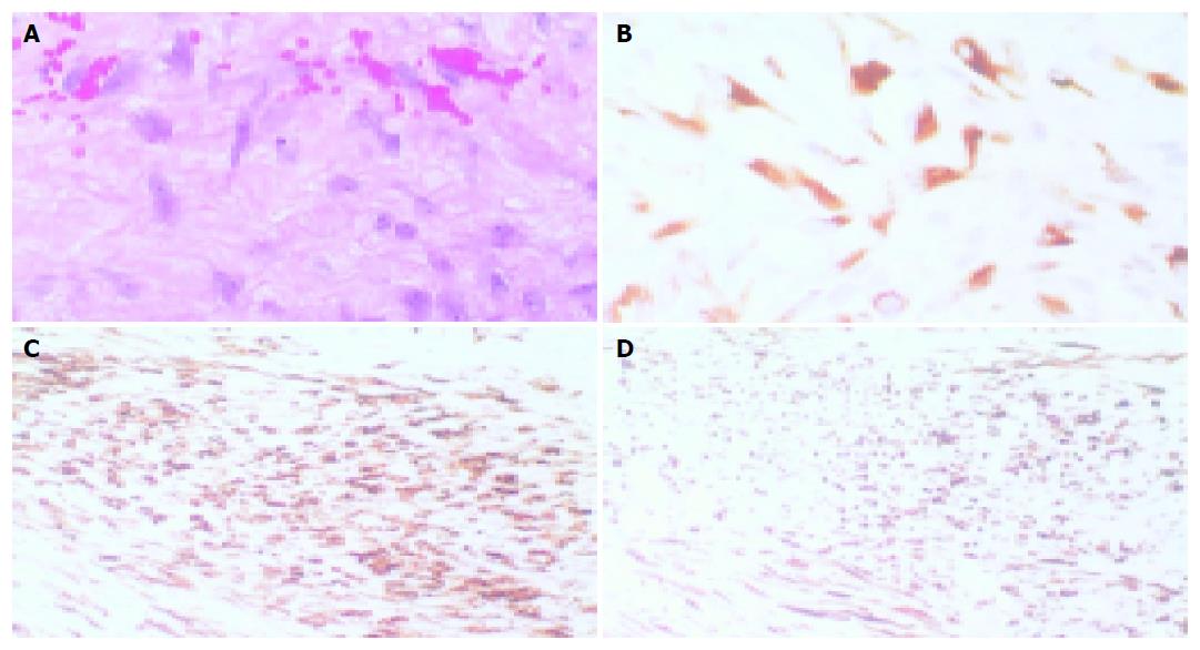
Figure 3 The tumoral lesion consisted of spindle cells growing in sweeping fascicles, with eosinophilic cytoplasm and sometimes plump nuclei (A, H&E 400×).
Most of the tumor cells show immunoreactivity for c-kit (CD117) in their cytoplasm (B, IHC 400×). Immunohistochemical stain for smooth muscle actin (C, IHC 200×) and for desmin (D, IHC 200×) is also present in some hypercellular areas of the lesion.
- Citation: Colombo P, Rahal D, Grizzi F, Quagliuolo V, Roncalli M. Localized intra-abdominal fibromatosis of the small bowel mimicking a gastrointestinal stromal tumor: A case report. World J Gastroenterol 2005; 11(33): 5226-5228
- URL: https://www.wjgnet.com/1007-9327/full/v11/i33/5226.htm
- DOI: https://dx.doi.org/10.3748/wjg.v11.i33.5226


