Copyright
©The Author(s) 2005.
World J Gastroenterol. Sep 7, 2005; 11(33): 5169-5173
Published online Sep 7, 2005. doi: 10.3748/wjg.v11.i33.5169
Published online Sep 7, 2005. doi: 10.3748/wjg.v11.i33.5169
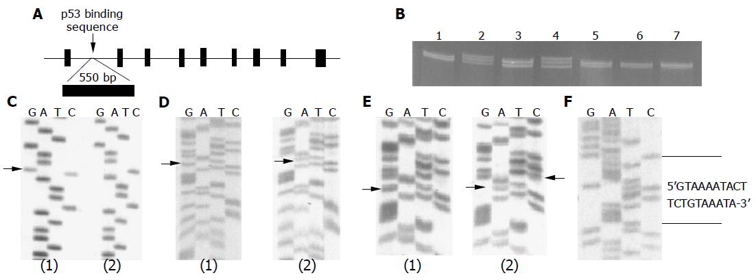
Figure 1 Structural feature and genetic variation analysis of p53R2.
A: A representative p53R2 gene structure including a regulatory region with p53 binding sequence within intron 1; B: representative cold SSCP result. p53R2 Regulatory region was amplified, and PCR products were run on a 20% SSCP gel at 15 °C. Lanes 2 and 4 are the heterozygous polymorphism pattern; C: alterations in regulatory region of p53R2 gene: (1) DdeI G/C, and (2) DdeI C/C genotypes; D: Alterations in regulatory region of p53R2 gene: (1) NdeI G/G and (2) A/A genotypes; E: Alterations in regulatory region of p53R2 gene: (1) NdeI G/G and HhaI T/T, and (2) NdeI A/A and HhaI T/C genotypes; F: Alterations in regulatory region of p53R2 gene: 20 bp insertion which replaced an ATTTT between nt 1 831 and nt 1 835.
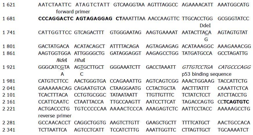
Figure 2 Representation of p53R2 intron 1.
The human p53R2 partial intron 1, as enumerated in the original description of the p53R2 gene (GenBank accession no.: NC000008), is shown. The three variable loci are underlined, boxed 5 bp is replaced by 20 bp nucleotides (5’-GTAAAATACTTCTGTAAATA-3’), the p53 binding sequence is italicized, and forward and reverse primers set in bold.
- Citation: Deng ZL, Xie DW, Bostick RM, Miao XJ, Gong YL, Zhang JH, Wargovich MJ. Novel genetic variations of the p53R2 gene in patients with colorectal adenoma and controls. World J Gastroenterol 2005; 11(33): 5169-5173
- URL: https://www.wjgnet.com/1007-9327/full/v11/i33/5169.htm
- DOI: https://dx.doi.org/10.3748/wjg.v11.i33.5169


