Copyright
©The Author(s) 2005.
World J Gastroenterol. Aug 14, 2005; 11(30): 4661-4666
Published online Aug 14, 2005. doi: 10.3748/wjg.v11.i30.4661
Published online Aug 14, 2005. doi: 10.3748/wjg.v11.i30.4661
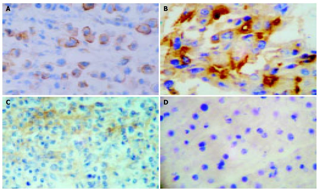
Figure 1 Characterization of MUC1 expression in PLC and cirrhotic liver tissues as well as normal liver tissues by immunohistochemical staining.
A: Overexpression of MUC1 on cell membranes (×400); B: positive staining of MUC1 in cytoplasm (×400); C: MUC1 weak expression in cirrhotic liver tissues (×400); D: MUC1 negative expression in normal liver tissues (×400).
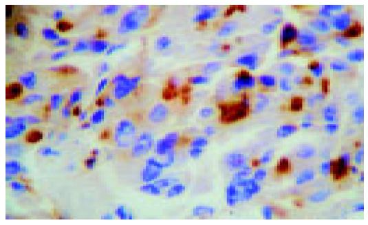
Figure 2 Positive expression of MUC1 protein in HCC samples (×400).
- Citation: Yuan SF, Li KZ, Wang L, Dou KF, Yan Z, Han W, Zhang YQ. Expression of MUC1 and its significance in hepatocellular and cholangiocarcinoma tissue. World J Gastroenterol 2005; 11(30): 4661-4666
- URL: https://www.wjgnet.com/1007-9327/full/v11/i30/4661.htm
- DOI: https://dx.doi.org/10.3748/wjg.v11.i30.4661


