Copyright
©The Author(s) 2005.
World J Gastroenterol. Aug 14, 2005; 11(30): 4644-4649
Published online Aug 14, 2005. doi: 10.3748/wjg.v11.i30.4644
Published online Aug 14, 2005. doi: 10.3748/wjg.v11.i30.4644
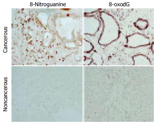
Figure 1 Localization of 8-oxodG and 8-nitroguanine in cancerous and noncancerous liver tissues in an ICC patient with a well-differentiated adenocarcinoma.
Formation of 8-oxodG and 8-nitroguanine was assessed by immunohistochemistry using an immunoperoxidase method. Paraffin sections (6 μm thickness) were incubated with a rabbit polyclonal anti-8-nitroguanine antibody and a mouse monoclonal anti-8-oxodG antibody. The original magnification is 200×.
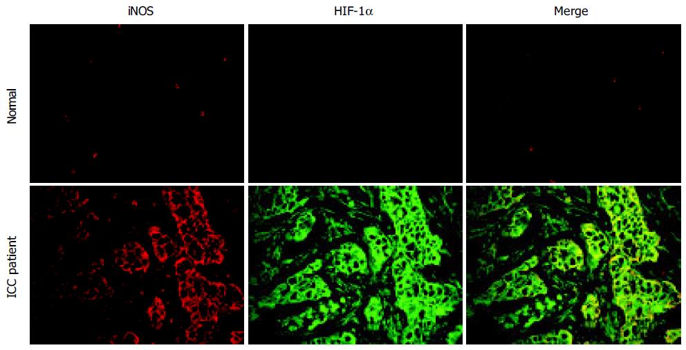
Figure 2 Localization of iNOS and HIF-1α in cancerous liver tissues in an ICC patient with poorly-differentiated adenocarcinoma.
The expression of iNOS and HIF-1α was assessed by using double immunofluorescence technique. Paraffin sections were treated with anti-iNOS and anti-HIF-1α antibodies. The original magnification is 400×.
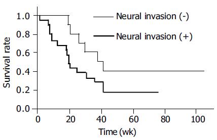
Figure 3 Overall survival curve of ICC patients.
Thin line, neural invasion evaluated as -; thick line, neural invasion evaluated as + (Kaplan-Meier method).
- Citation: Pinlaor S, Sripa B, Ma N, Hiraku Y, Yongvanit P, Wongkham S, Pairojkul C, Bhudhisawasdi V, Oikawa S, Murata M, Semba R, Kawanishi S. Nitrative and oxidative DNA damage in intrahepatic cholangiocarcinoma patients in relation to tumor invasion. World J Gastroenterol 2005; 11(30): 4644-4649
- URL: https://www.wjgnet.com/1007-9327/full/v11/i30/4644.htm
- DOI: https://dx.doi.org/10.3748/wjg.v11.i30.4644


