Copyright
©The Author(s) 2005.
World J Gastroenterol. Aug 7, 2005; 11(29): 4505-4510
Published online Aug 7, 2005. doi: 10.3748/wjg.v11.i29.4505
Published online Aug 7, 2005. doi: 10.3748/wjg.v11.i29.4505
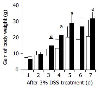
Figure 1 Changes in body weight in normal serum- (open columns), anti-ECP- (closed columns) treated rats during DSS treatment.
The body weight gain after DSS treatment was significantly greater in anti-ECP-treated rats compared with normal serum-treated rats. Rats treated with the antibodies or normal serum intraperitoneally were also treated with 3% DSS in drinking water for 7 d (n = 7, aP<0.05 vs DSS+normal serum).
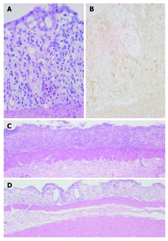
Figure 2 Colonic mucosa at 3 d post-DSS treatment showing eosinophils in the lamina propria.
HE staining (A) and immunostaining for ECP (B) as described under Materials and methods. ECP-positive cells (brown) appear in close proximity to damaged crypts in the lamina propria and partially in the extracellular interstitium of DSS-induced colitis. Original magnification, ×400. Colonic mucosa at 7-d treatment of DSS. (C) Colonic ulceration in normal serum-treated rats. (D) Reduced severity of colonic mucosal ulceration in anti-ECP-treated rats. HE stain. Original magnification, ×100.
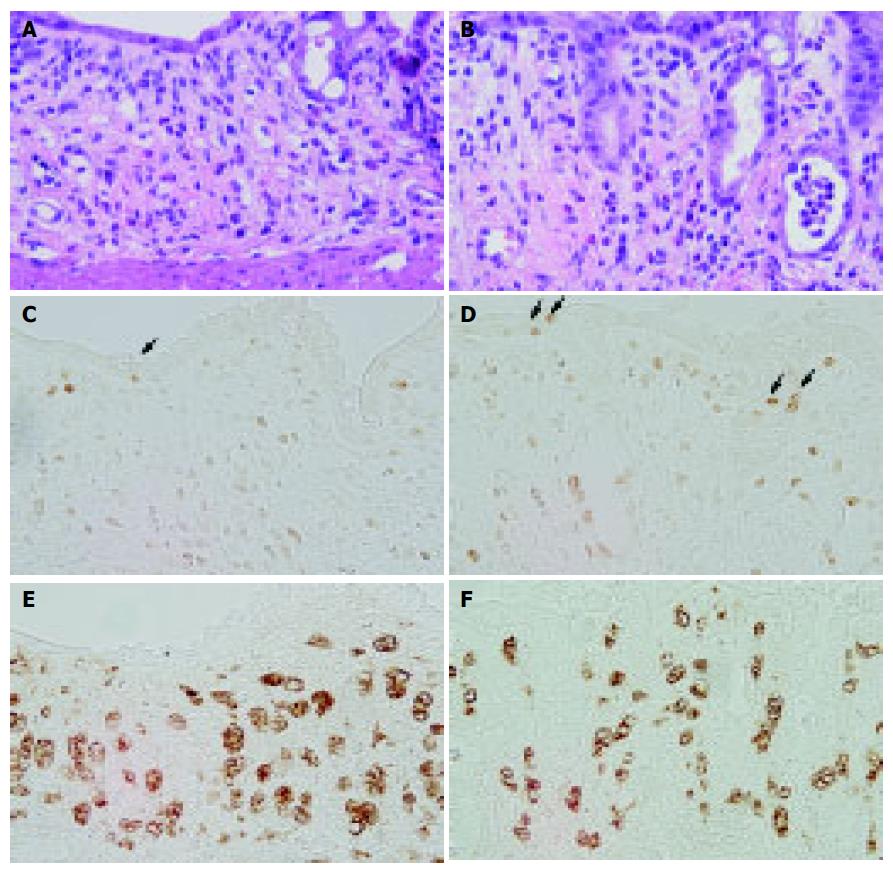
Figure 3 Colonic mucosa in normal serum-treated rats (A, C, and E) and anti-ECP1-treated rats (B, D, and F) at 7-d treatment of DSS.
HE staining (A and B), immunostaining for Ki-67 (C and D) and ED1 (E and F) as described under Materials and methods. Treatment with ECP antibody increased the number of Ki-67-positive cells in the regenerated surface epithelium and reduced the size of activated macrophages present in the lamina propria. Original magnification, ×400.
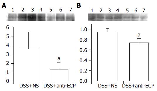
Figure 4 Detection of ED1 (A) and MIF (B) by Western blot analysis in colonic tissues after 7 d of DDS treatment in normal serum-treated rats (lanes 1-4) and anti-ECP-treated rats (lanes 5-7).
The colonic tissue was collected from the lesion area and examined by Western blot analysis as described under Materials and methods. The relative expression levels of ED1 and MIF in the damaged colonic tissue were significantly lower in anti-ECP-treated rats than in normal serum-treated rats. The relative protein expression was quantified by densitometric analysis (n = 7, aP<0.05 vs DSS+normal serum).
- Citation: Shichijo K, Makiyama K, Wen CY, Matsuu M, Nakayama T, Nakashima M, Ihara M, Sekine I. Antibody to eosinophil cationic protein suppresses dextran sulfate sodium-induced colitis in rats. World J Gastroenterol 2005; 11(29): 4505-4510
- URL: https://www.wjgnet.com/1007-9327/full/v11/i29/4505.htm
- DOI: https://dx.doi.org/10.3748/wjg.v11.i29.4505


